Figure 2. Comparison of primary sphere
formation by rabbit CEC from the periphery and center of the cornea. A:
The
number of spheres obtained from limbal CEC (n=10) was significantly
higher than that from central CEC (n=10) after 7 days of culture. The
experiment was repeated twice and representative data are shown. The
asterisk indicates a p=0.0028 (unpaired t-test). B: The
mean sphere size exceeded 250 μm on day 7.
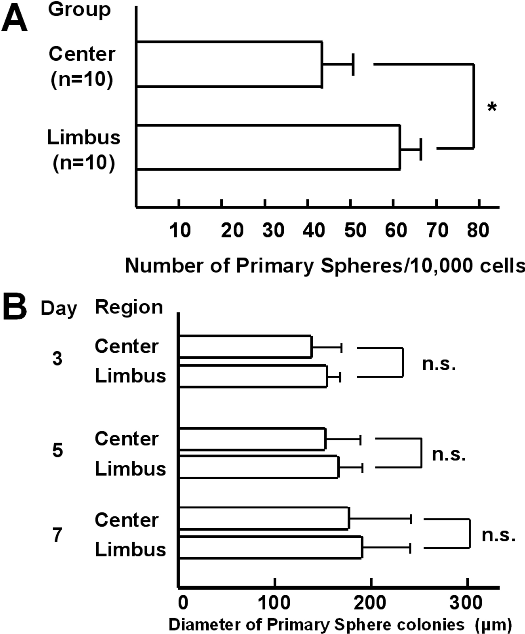
 Figure 2 of Mimura, Mol Vis 2010; 16:1712-1719.