Figure 3. Summary of FOXE3 mutations. A: Alignment of FOXE3 proteins showing region of p.Tyr191-Pro193 duplication. Please note two YAP amino acid motifs present
in normal human and cow FOXE3 proteins and an additional insertion of this motif identified in Patient 2. B: Schematic drawing of FOXE3 protein with human mutations. Forkhead domain is shown in dark gray. Mutations resulting in recessive
phenotypes are indicated with black arrows while mutations causing dominant disease- with blue arrows. The variant identified
in Patient 2 is indicated with a gray arrowhead below the protein. Recurrent mutations are denoted with extended arrows. Positions
of mutations identified in this study are shown with asterisks.
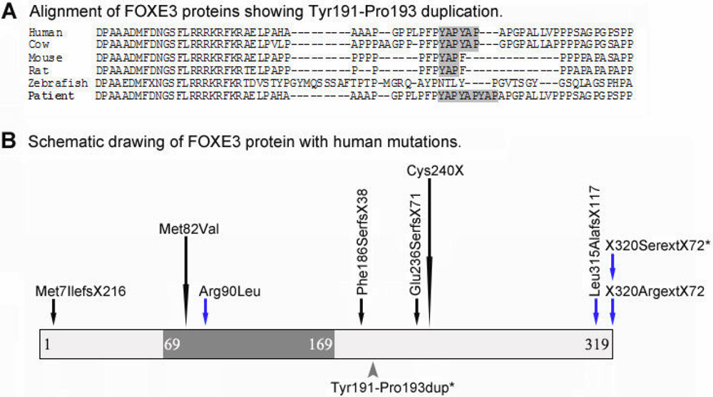
 Figure 3 of
Brémond-Gignac, Mol Vis 2010; 16:1705-1711.
Figure 3 of
Brémond-Gignac, Mol Vis 2010; 16:1705-1711.