Figure 2. Evaluation of limbal stem cell and corneal differentiation markers pre and post air lifting in cultured limbal cells on AM
and PD groups. A: Representative fluorescent staining profile shows that P63, a putative stemness marker, expressed in nucleus and K3, a differentiation
marker, in cytoplasm and Cx43 in cell membrane and near the nucleus in cultured cells on amniotic membrane (AM) and plastic
dish (PD) before and after air lifting. The nuclei were stained by Dappi (Blue). Scale bar: 100 µm. B: Comparative data for PD and AM group for P63, K3, and CX43, before and after air lifting by flowcytometry analysis. C: Immuno staining of cornea and limbal tissues for P63 and K3 Marker. Scale bar: 100 µm. D: RT–PCR shows the gene expression of P63,K3, ABCG2 and GAPDH as internal control before and after air lifting in AM, PD as
well as limbal and corneal cells. Results were presented as percentage of 3 to 4 different experiments and expressed as mean±SD.
The asterisk indicates a p<0.05 and the double asterisk indicates a p<0.01.
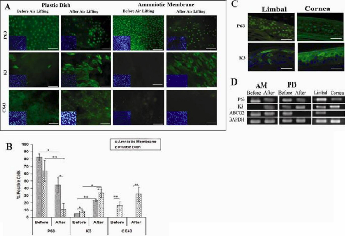
 Figure 2 of
Ebrahimi, Mol Vis 2010; 16:1680-1688.
Figure 2 of
Ebrahimi, Mol Vis 2010; 16:1680-1688.