Figure 1. Human limbal explants expanded on human amniotic membrane (HAM) and plastic dish (PD) for 14 days, followed by air lifting
for 16 days. A: Phase-contrast microscopy of the cells shows that cells on HAM are small and compact compared with those in PD pre air lifting;
however, the morphology of the cells in the PD group changes to squamous epithelial cells post air lifting. Scale bar: 100
µm. B: Assessment of cell heterogeneity in R1: 10–200 or limbal stem cell (LSC), R2: 200–400 or Transient amplifying cells (TAC),
and R3: >400 or terminal differentiated cells (TDC) by light scatter showed that cells in PD are more differentiated than
those in AM. C: The comparison of cells in PD and AM groups before and after air lifting shows cells differentiated to large cells with
high granularity. Results were presented as percentage of 3 different experiments and expressed as mean±SD. The astereisk
indicates a p<0.05 and the double asterisk indicates a p<0.01.
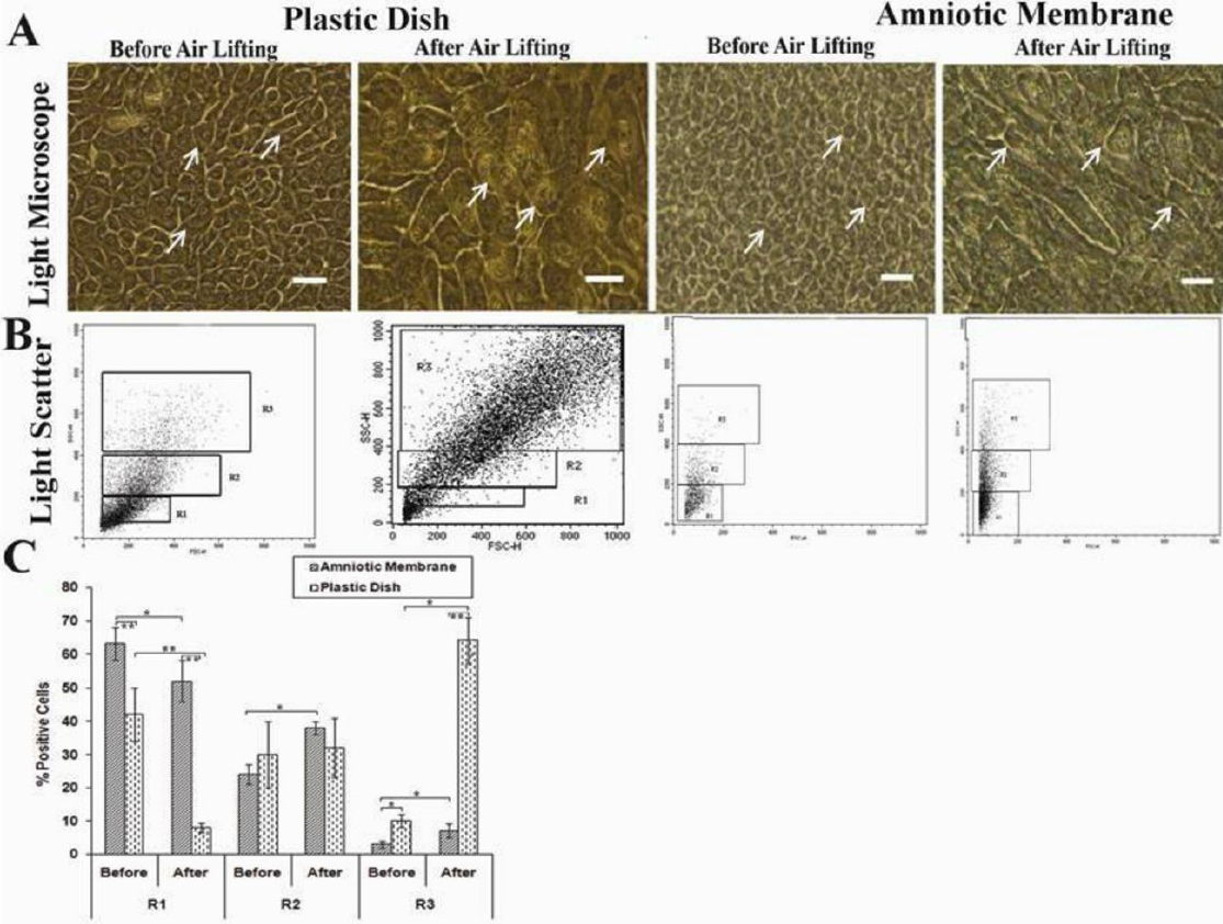
 Figure 1 of
Ebrahimi, Mol Vis 2010; 16:1680-1688.
Figure 1 of
Ebrahimi, Mol Vis 2010; 16:1680-1688.