Figure 4. Retinal structure. A-D: Retinal structure is revealed by representative photomicrographs from the central superior retina from Fat1+/VPP+, Fat1–/VPP+,
Fat1+/VPP–, and Fat1–/VPP– mice at 4 weeks of age. E-H: Photomicrographs at 16 weeks of age are shown. I-L: Photomicrographs at 28 weeks of age are shown. Magnification bar equal to 50 microns.
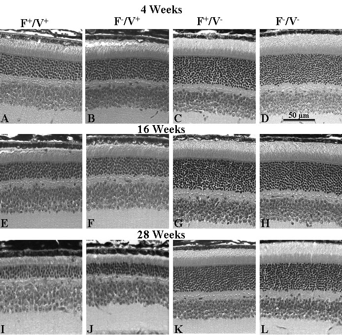
 Figure 4 of
Li, Mol Vis 2010; 16:1669-1679.
Figure 4 of
Li, Mol Vis 2010; 16:1669-1679.