Figure 2. Typical appearance of
kidney-shaped and biconvex lenses. Panels A and B each
show a single “slice” from the central region of a magnetic resonance
imaging (MRI) scan: a nonkidney- shaped lens (A) and a
kidney-shaped lens (B). Panels C and D show
MRI3dX mesh models of the lenses from two eyes: a lens with a normal
biconvex appearance (C) and a lens showing a depression the
anterior surface, characteristic of kidney-shaped lenses (D).
The scale bar in panel D is only an approximation since the
true dimensions are altered due to the presentation in perspective.
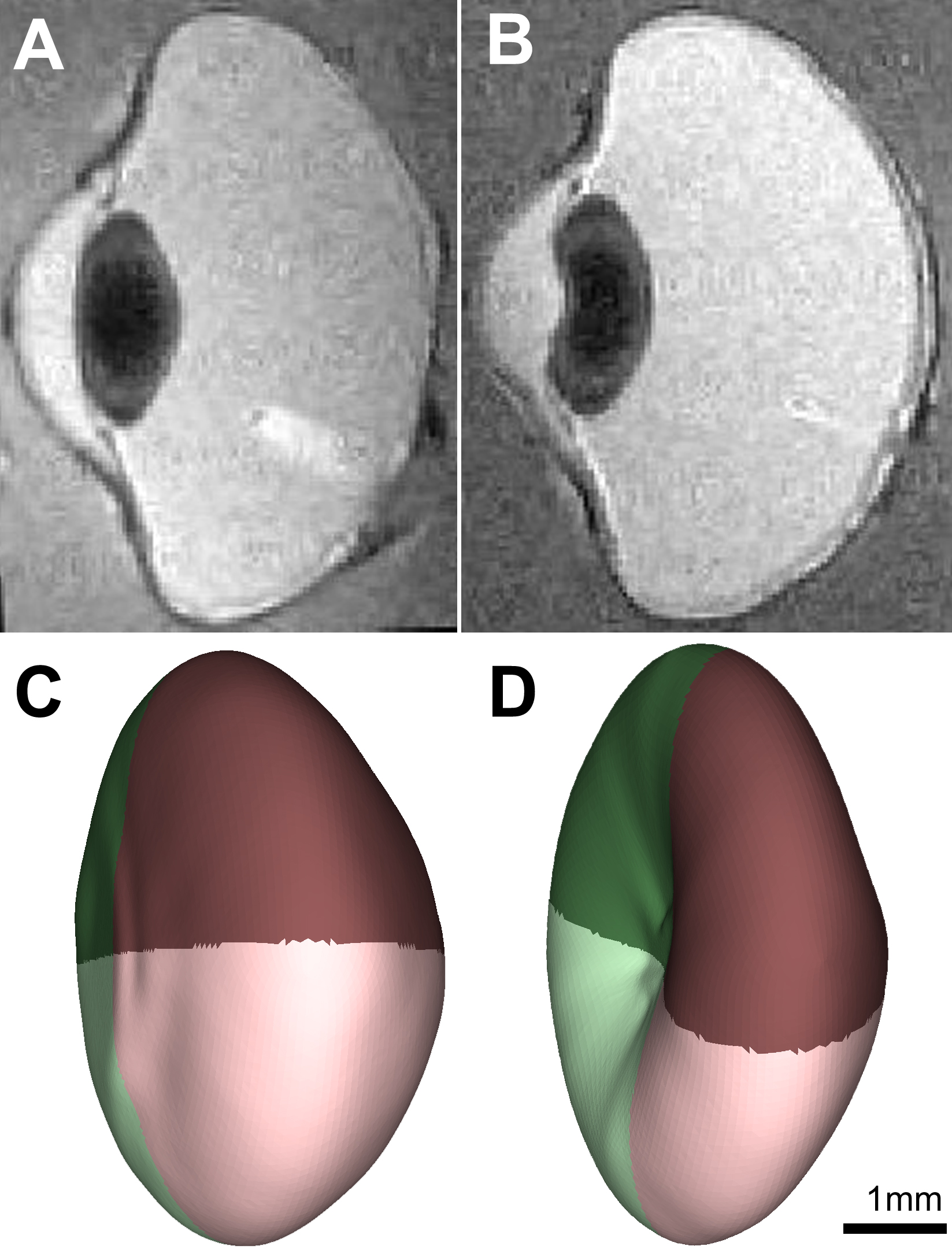
 Figure 2 of Tattersall, Mol Vis 2010; 16:144-153.