Figure 1. Arrangement of eyes for magnetic
resonance imaging scanning. The figure shows 16 paraformaldehyde-fixed
chicken eyes embedded in low-melting-point agarose in a 2×2×4 array in
readiness for an overnight magnetic resonance imaging (MRI) scan. Note
that the eye in the lower-right quadrant of the uppermost layer was
positioned in an inverted orientation to permit unambiguous
identification of each eye in the resultant MRI image. In the remaining
three eyes of the uppermost layer, it is possible to see the ink mark
on the nasal cornea, which was used to indicate the original
orientation of the eye in the head.
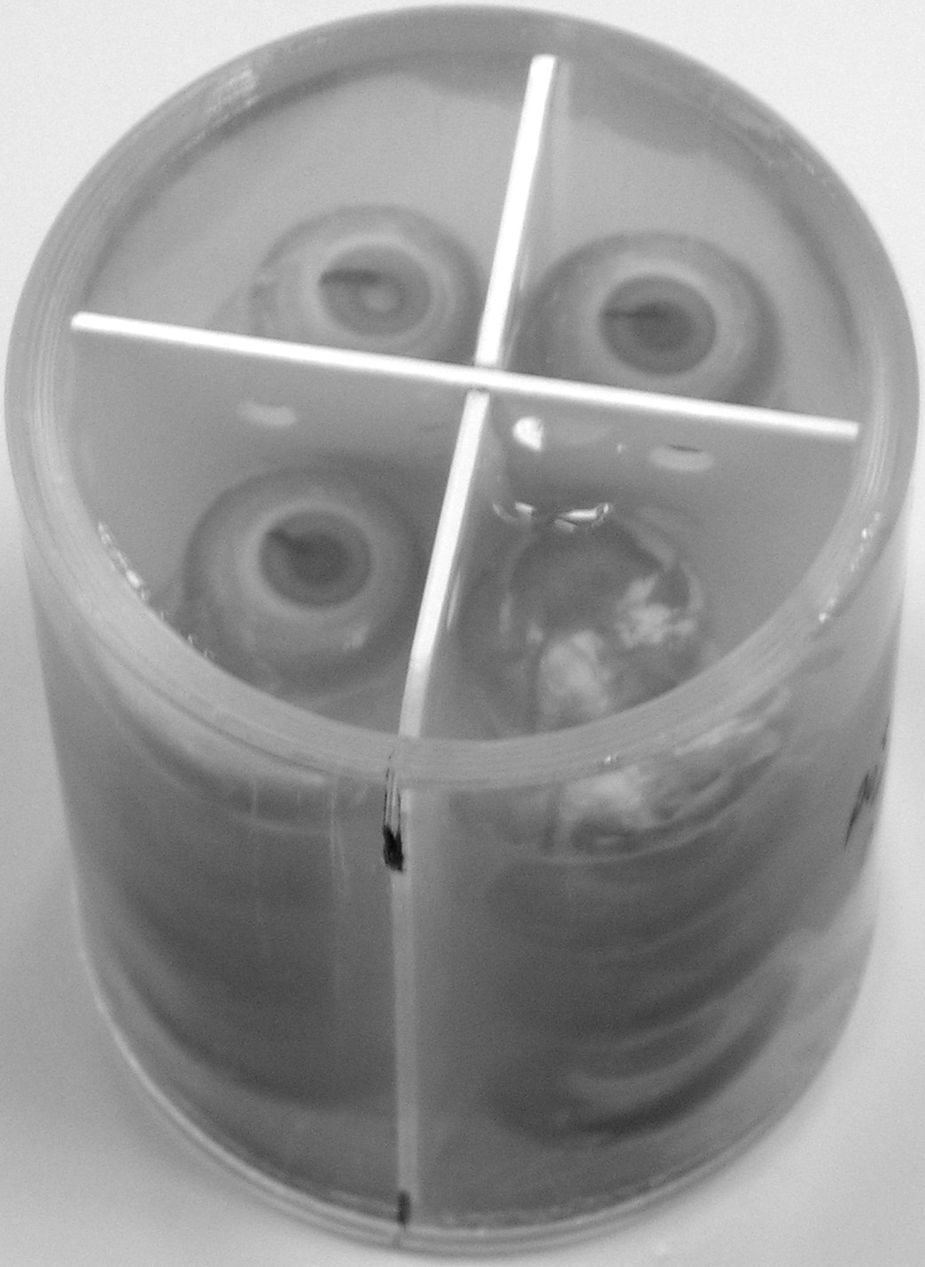
 Figure 1 of Tattersall, Mol Vis 2010; 16:144-153.