Figure 2. HPLC chromatograms of plasma mangiferin. HPLC separation was performed on plasma samples using the Agilent 1200 Series Rapid
Resolution system. A COSMOSIL 5C18—MS—IIanalytical column (4.6 mm×250 mm,5 μm) was used and operated at 25 °C. The mobile phase consisted of methanol −2% glacial
acetic acid (40:60 v:v). Typical chromatograms of blank plasma, blank plasma spiked with mangiferin and I.S., and rat plasma
sample after injection of mangiferin are presented. Mangiferin and the I.S. were eluted at 5.6 and 12.16 min, respectively.
The total run time was less than 30 min. A good separation of the I.S. and mangiferin was obtained under the specified chromatographic
conditions. There is no disturbance from the background signals in the plasma after the protein precipitation step. A: Typical chromatogram of blank plasma. B. Typical chromatogram of blank plasma spiked with standard mangiferin (5 μg/ml) and I.S. panel. C. Typical chromatogram of blood sample containing mangiferin (24.14 µg/ml) collected at 0.5 h after mangiferin administration
(10 mg/kg, i.v.). D: Typical chromatogram of blood sample containing mangiferin (73.88 µg/ml) collected at 0.5 h after mangiferin administration
(25 mg/kg, i.v.). E: Typical chromatogram of blood sample containing mangiferin (221.54 µg/ml) collected at 0.5 h after mangiferin administration
(50 mg/kg, i.v.).
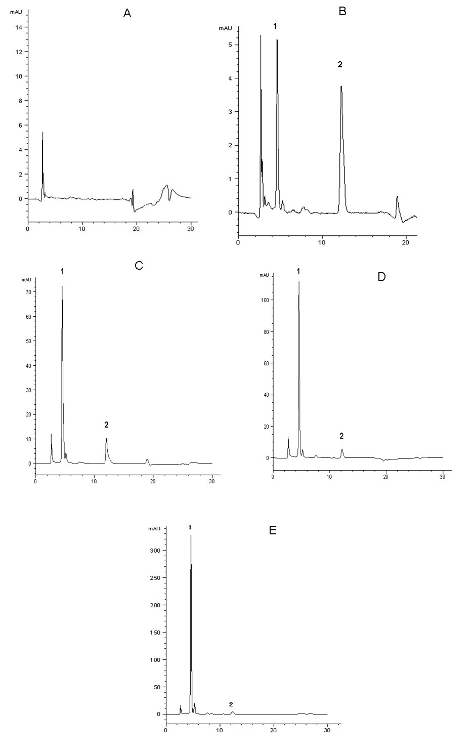
 Figure 2 of
Hou, Mol Vis 2010; 16:1659-1668.
Figure 2 of
Hou, Mol Vis 2010; 16:1659-1668.