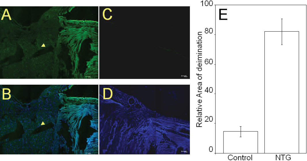
Figure 4. Representative immunohistochemical analyses of NTG (70 F) and control (72F) donor optic nerve sections.
A: Immunoreactivity for protein-bound citrulline detected by a rabbit polyclonal antibody to citrulline after monoxime modification
as described in methods.
B: Merged image of anti-citrulline with DAPI.
C: Control donor section probed for immunoreacitivty with anti-citrulline.
D: Merged image of anti-citrulline and DAPI for control donor section.
E: Densitometric analysis of immunohistochemical detection of protein-bound citrulline in NTG eyes. The data was analyzed using
Image J software.
 Figure 4 of
Cafaro, Mol Vis 2010; 16:1654-1658.
Figure 4 of
Cafaro, Mol Vis 2010; 16:1654-1658.