Figure 2. Western blotting was detected of proliferating cell nuclear antigen (PCNA) in rLEC. PCNA, and β-actin were determined in rLECs
by western blotting (A). The blot was stripped reprobed with anti-β-actin as an internal control: Lanes M, 1, 2, 3, 4 and 5 are protein marker,
normal control, 0.1 ng/ml for 24 h, 0.1 ng/ml rapamycin for 48 h, 10 ng/ml rapamycin for 24 h, and 10 ng/ml rapamycin for
48h, respectively. The histogram (B) summarizes the results of scanning densitometry of PCNA in the blot. Data were standardized by an internal loading control
(β-actin). All differences were statistically significant, *p≤0.05.
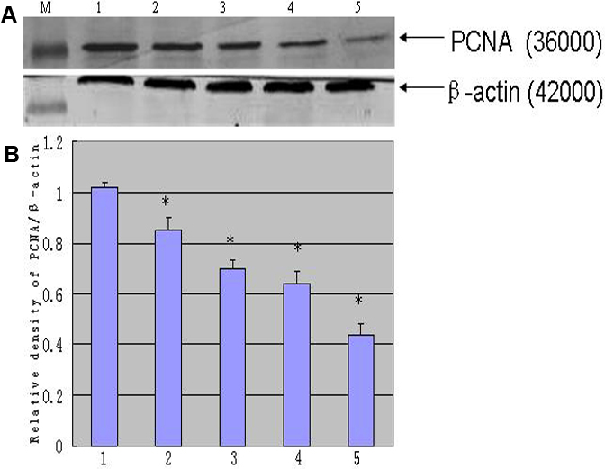
 Figure 2 of
Liu, Mol Vis 2010; 16:1646-1653.
Figure 2 of
Liu, Mol Vis 2010; 16:1646-1653.