Figure 2. Location of the Leu113Ser mutation in the crystal structure of the NT-4-TrkB complex. The two chains of the dimeric NT-4 are
in cyan and blue, respectively. The alpha carbon of the mutated residue is shown as a magenta sphere, with the prime symbol
denoting the residue of the second subunit. TrkB domains are shown in orange and red color. The Leu113 residue is located
close to the TrkB binding site.
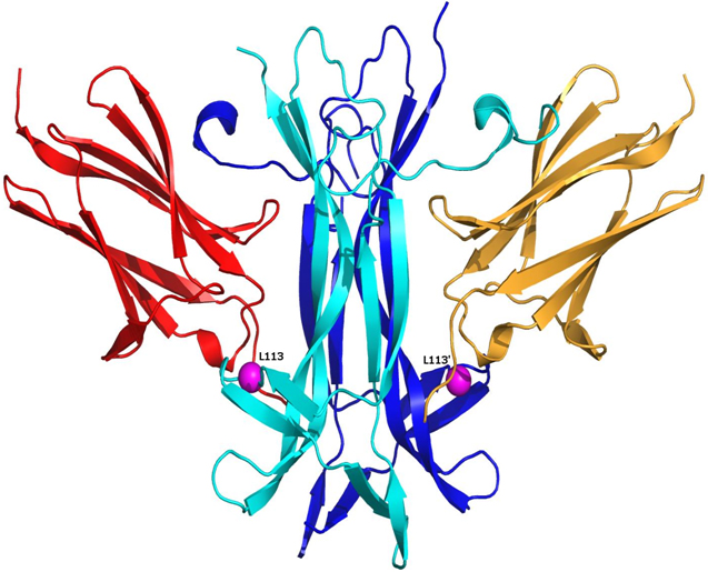
 Figure 2 of
Vithana, Mol Vis 2010; 16:1640-1645.
Figure 2 of
Vithana, Mol Vis 2010; 16:1640-1645.