Figure 2. Fundus photographs and
electroretinography results of the proband with X-linked retinitis
pigmentosa,. The bone spicule pigmentation and attenuated retinal
vessels can be seen bilaterally. Both the rod and cone
electroretinogram amplitudes were below 10% of normal
electroretinography (ERG; norms for scotopic [skot] ERG: b-wave 27.6
μV±5.2; norms for photopic [phot] ERG: b-wave 70 μV±8.9).
Abbreviations: div means division.
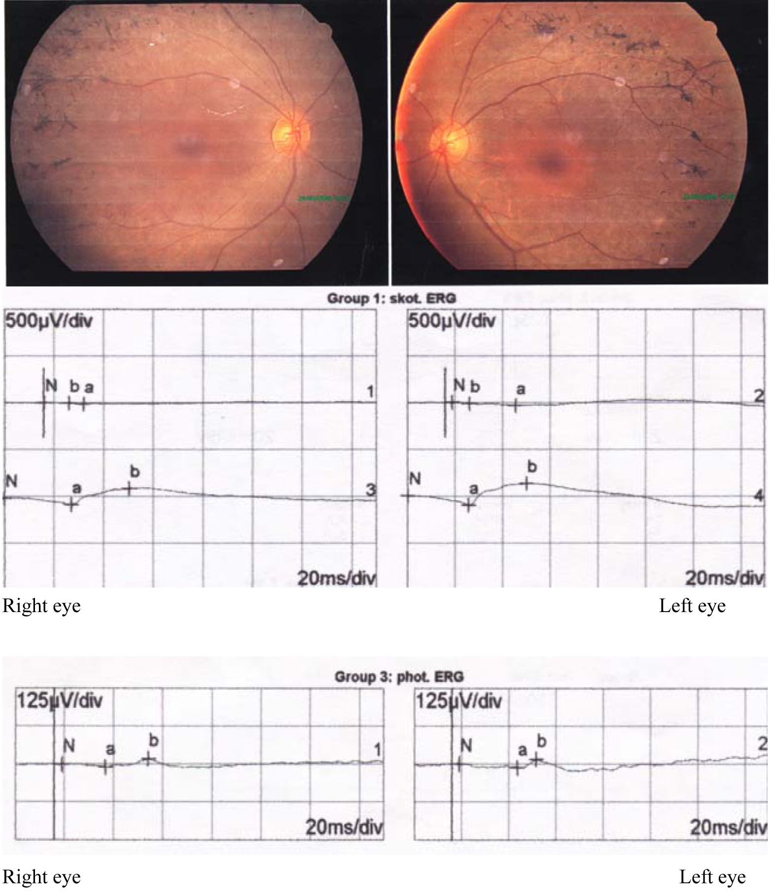
 Figure 2 of Sheng, Mol Vis 2010; 16:1620-1628.