Figure 4. A: Magnified images of the immunolocalization of anti-aquaporin water channels (AQP4) and anti-Kir4.1 antibodies in the inner
chick retina, demonstrating differences in the spatial distribution of AQP4 and Kir4.1 (Upper panels show confocal images
illustrating the immunodistribution of AQP4 and Kir4.1 channel expression in normal chick retina. Lower panels show the associated
differential interference contrast images). AQP4 expression was prominent in the nerve fiber layer (NFL), ganglion cell layer
(GCL), and inner plexiform layer (IPL), whereas, Kir4.1 expression was more prominent in the IPL with some diffuse staining
evident in the NFL/GCL and branching processes extending out into the inner nuclear layer (INL). The scale bar represents
50 µm. B: AQP4 expression following 2 days of lens wear. Upper images- Confocal microscopy showing AQP4 labeling and the accompanying
differential interference contrast images from the myopic (left) and fellow eye (right) 2 days post lensing. Lower images
- Similar confocal and interference contrast images of AQP4 labeling in hyperopic and fellow eyes. The scale bar represents
50 µm. C: Kir4.1 immunoreactivity after 4 days of lens wear. Upper panels show Kir4.1 immunopositivity in the myopic and fellow eye.
Lower panels show Kir4.1 immunopositivity in the hyperopic eye and fellow eye. Note the appearance of strong labeling in the
IPL of the hyperopic eye compared to the fellow eye below and lesser staining of Kir4.1 in the myopic eye compared to the
fellow eye. The scale bar represents 50 µm.
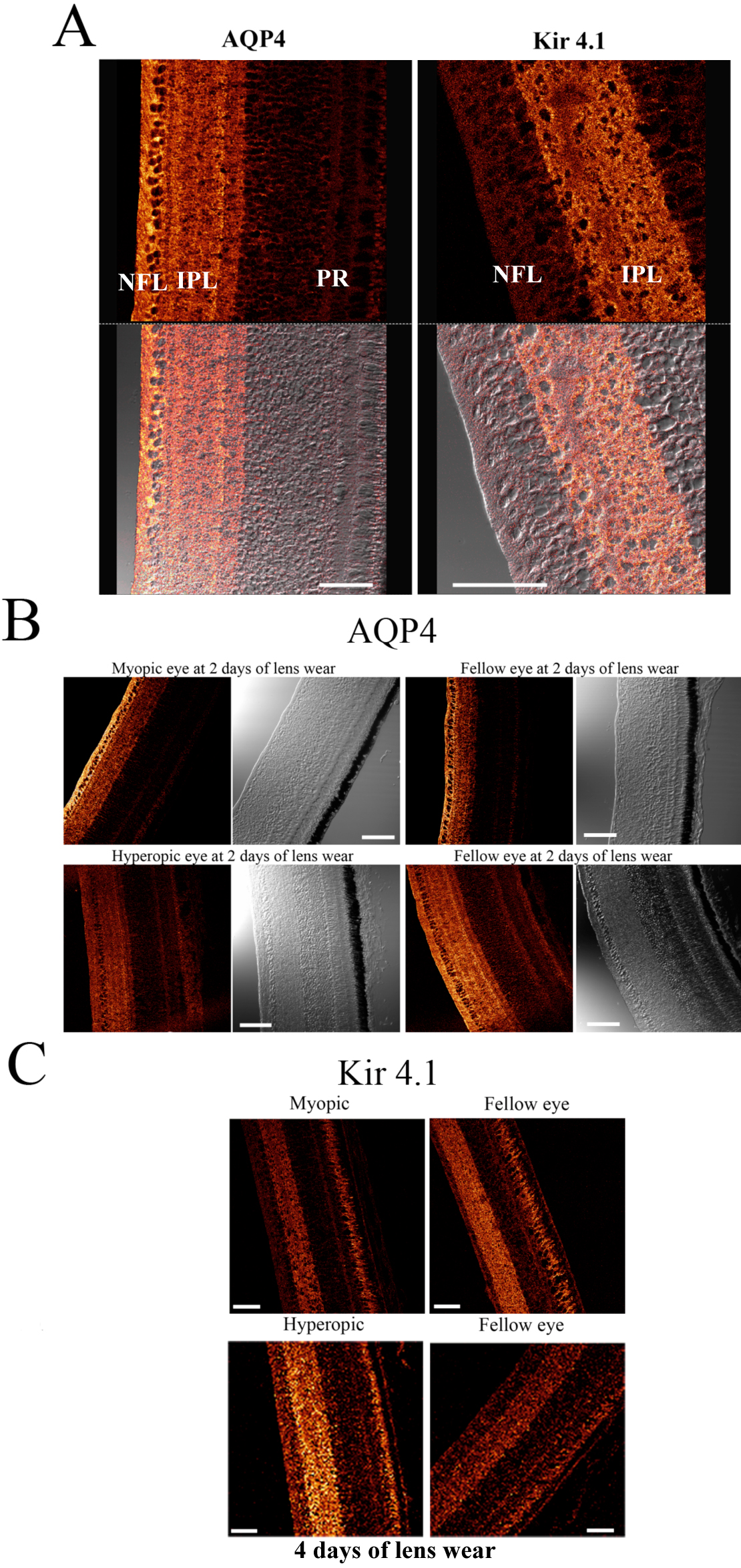
 Figure 4 of
Goodyear, Mol Vis 2010; 16:1610-1619.
Figure 4 of
Goodyear, Mol Vis 2010; 16:1610-1619.