Figure 3. Immunolocalization of aquaporin water channel AQP4, inward rectifying potassium Kir4.1 channel, and glial fibrillary acidic
protein (GFAP) expression in the chick retina. Staining of AQP4 (red) and Kir4.1 (green) is similar to that of GFAP (purple),
a known marker of Muller cell processes. The layers of the retina are indicated as NFL for nerve fiber layer, IPL for inner
plexiform layer and PR for photoreceptor layer.
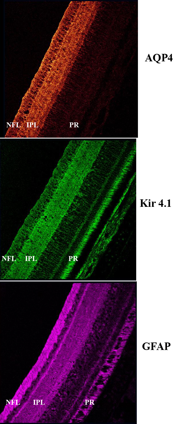
 Figure 3 of
Goodyear, Mol Vis 2010; 16:1610-1619.
Figure 3 of
Goodyear, Mol Vis 2010; 16:1610-1619.