Figure 2. Photographs of the cornea from
six individuals examined using slit lamp examination. Slit lamp
photographs of patient V-19 of Family One at 27 years of age show
opacities in the central stroma and linear forms in the left cornea (A
and B; A: OD and B: OS). The image of case
III-1 shows irregularity of the epithelial surface with subepithelial
and anterior stromal scarring in the left eye (C). The image of
case IV-10 revealed the presence of a network of linear opacities
associated with polymorphic anterior stromal opacities in the right eye
(D). The image of case IV-13 at 50 years of age shows opacities
in the central stroma and linear forms in the left cornea (E).
The image of case II-1 of Family Two at age 25 shows a network of
linear opacities associated with other smaller opaque spots and
refractile lattice lines in the left eye (F) and the right eye
shows penetrating keratoplasty with characteristic mydriasis of
Urrets-Zavalia syndrome (G). The photographs of case I-1 of
Family Three at 52 years of age show thick lattice lines and yellowish
discoloration in the anterior stroma, resulting in clouding of the
central cornea (H and I).
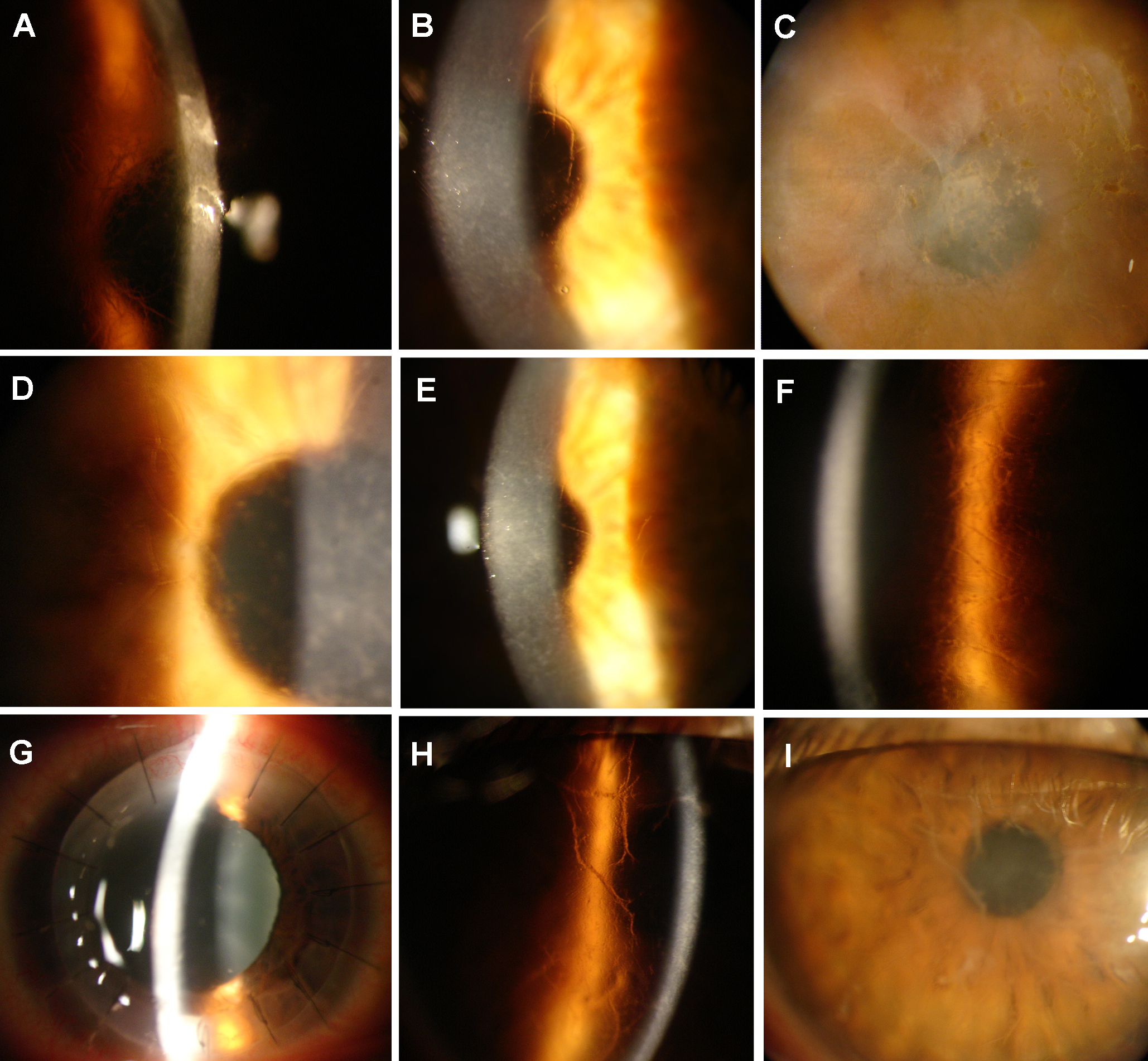
 Figure 2 of Romero, Mol Vis 2010; 16:1601-1609.