Figure 3. Fundus photographs for the elder brother (III-13) of the proband. Panel A describes the right eye showing a foveal stellate cystic change and a peripheral retinoschisis in the upper temporal retina.
Panel B describes the left eye showing peripheral retinoschisis involving the macula.
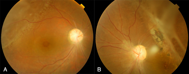
 Figure 3 of
Xu, Mol Vis 2010; 16:1593-1600.
Figure 3 of
Xu, Mol Vis 2010; 16:1593-1600.