Figure 2. Photographs for the proband (III-14) of the family with X-linked juvenile retinoschisis. A: Fundus photograph of the right eye. B: Fundus photograph of the left eye. C: Slit-lamp photograph showing a membrane with blood vessels behind the lens. D: Ultrasonography of the right eye and left eye. E: Fundus photograph of the left eye after vitrectomy.
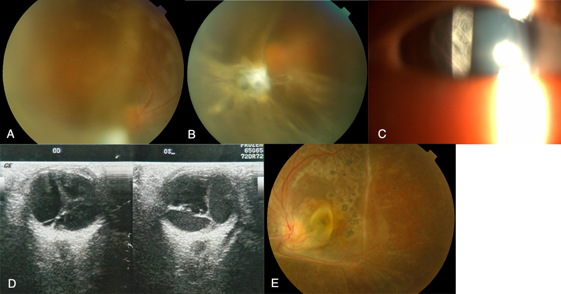
 Figure 2 of
Xu, Mol Vis 2010; 16:1593-1600.
Figure 2 of
Xu, Mol Vis 2010; 16:1593-1600.