Figure 4. Histopathological findings of
the corneal button taken from the right eye. A: Hematoxylin and
eosin staining shows amorphous deposits in the subepithelial region
(asterisk). The overlying epithelium is degenerated, and Bowman’s layer
is completely replaced by deposits. Underneath these amorphous
deposits, there are globular deposits of various sizes with irregular
peripheral margins that stained weakly with eosin (arrow). B:
Amyloidal deposition is confirmed in the subepithelium with Congo red
staining (asterisk). While the globular deposits of various sizes
located primarily in the anterior stroma stained negatively with Congo
red (arrow) (original magnification 100×).
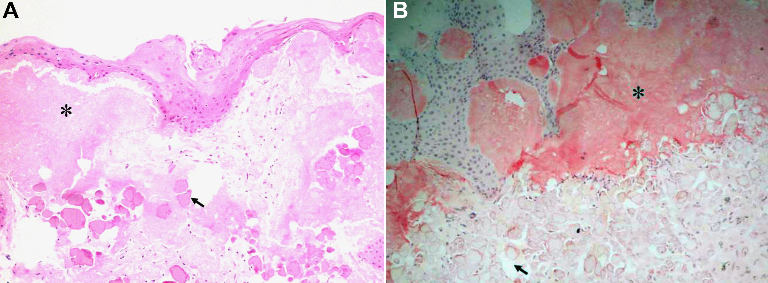
 Figure 4 of Zhang, Mol Vis 2010; 16:1570-1575.