Figure 3 of
Zhang, Mol Vis 2010; 16:1570-1575.
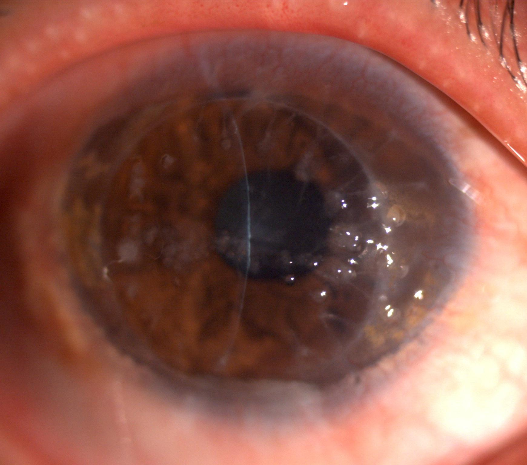
Figure 3.
Slitlamp photographs of the right cornea of the proband four year after penetrating keratoplasty. Elevated, mulberry-like gelatinous lesions companied with yellowish globular droplets are showed.
 Figure 3 of Zhang, Mol Vis 2010; 16:1570-1575.