Figure 2. Light- and electron-microscopic
retinal morphology in normal and canine cone-rod dystrophy 3 (crd3)
affected dogs. In the retina of a 27-weeks-old non-affected dog (A),
the
outer nuclear layer (ONL) comprises approximately 10 rows of rod
nuclei and a single distal row of cone nuclei. The inner and outer
segments of the photoreceptors (IS, OS) are of consistent proportions,
tightly aligned, and parallel, and the distal OS tips are in close
proximity to the apical membrane of the retinal pigment epithelium
(RPE). In retinas of 4.7- and 13.4-weeks-old crd3-affected dogs (B,
C), rod and cone IS and OS lack the tightly packed highly
parallel organization of a normal photoreceptor layer, and the distal
OS tips appear to be more distant from the RPE apical membrane than in
normal dogs. In the retina of an 18-weeks-old crd3-affected dog (D),
IS
and OS are disarrayed and disorganized, and a distinct gap is
observed between the RPE and the OS (arrows). The retinas of 26-weeks-
and 5 years-old crd3-affected dogs (E, F) exhibit
continued photoreceptor degeneration as evidenced by loss of cone and
rod IS, OS, and nuclei. Electron micrograph of the retina of a
27-weeks-old nonaffected dog (G) shows that the microvilli from
the RPE apical membrane extend to invest the photoreceptor OS. Electron
micrographs of the retina of a 13.4-weeks-old crd3-affected dog (H,
I) show that the RPE apical microvilli form a tangled flattened
mat that does not extend to invest the photoreceptor OS (arrows).
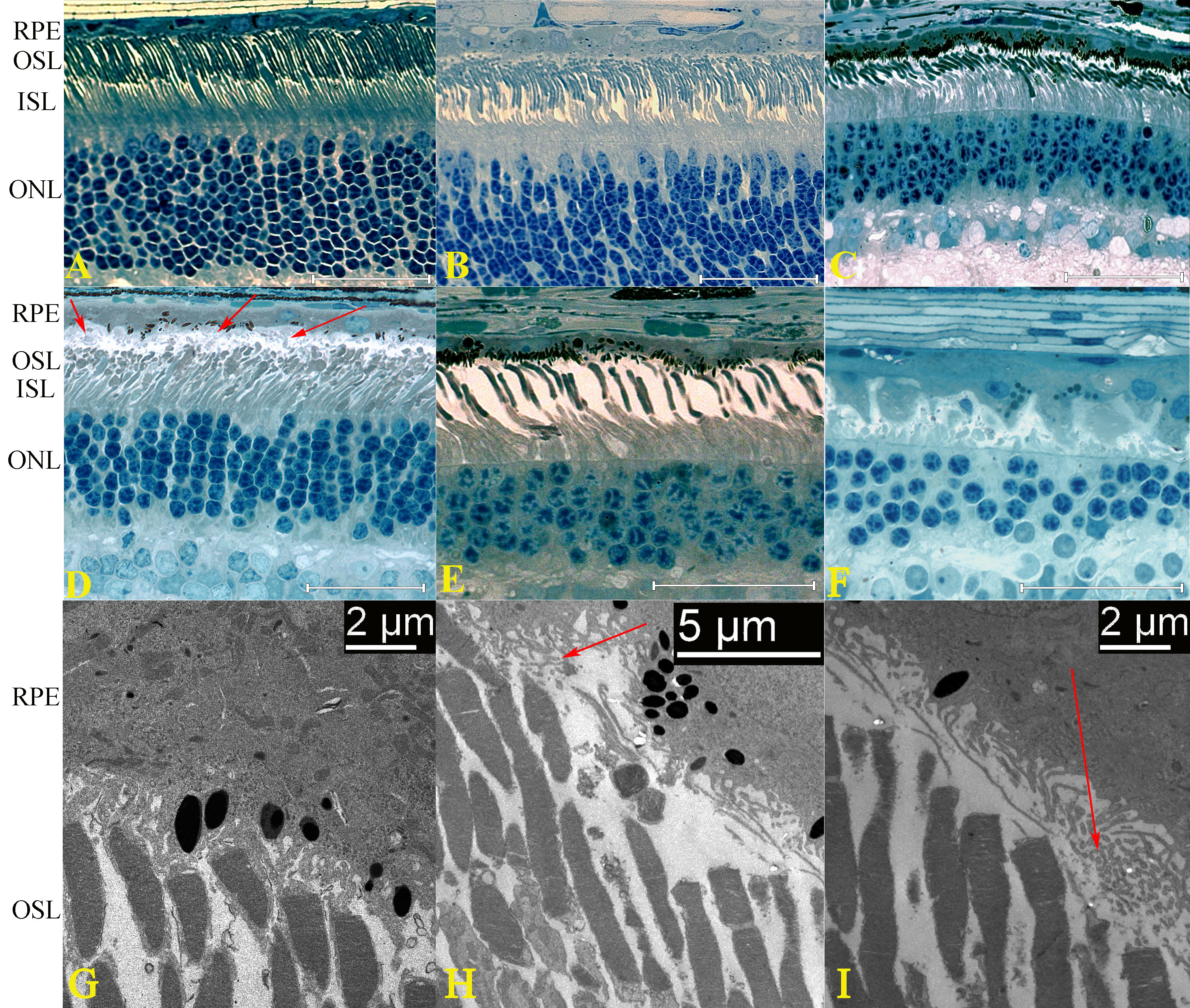
 Figure 2 of Goldstein, Mol Vis 2010; 16:1549-1569.