Figure 1. Electroretinograms of normal and
affected dogs. Electroretinograms (ERGs) from a 7 weeks old normal dog (A),
a
12
weeks
old
dog affected with canine cone-rod dystrophy 3 (crd3; B),
a
crd3-affected
dog aged 1.2 years (C), a 2 years old
crd3-affected dog (D), and a 4.9 years old crd3-affected dog (E).
Each
vertical
panel
presents
electroretinogram (ERG) responses to a
red flash, a blue flash, a white flash, 5 Hz low-intensity white
flashes
(Rod), and 30 Hz high-intensity white light flicker (Cone). Short
vertical arrows under the Rod and Cone flicker responses indicate the
onset of the flickering light stimuli. Red and
White traces represent mixed rod-cone responses, Blue and Rod traces
are rod-specific, and Cone traces are cone-specific. Responses of the
12-weeks-old crd3-affected dog appear normal (B), but by 15
months of age, cone dysfunction is detected as reduced 30 Hz flicker
responses (C), and is followed at later ages by continued
deterioration of both cone and rod responses (D, E). At
all ages, the loss of cone function is more prominent than that of
rods. Vertical calibration bar=100 µV; horizontal=200 ms for rod
flicker; and other responses are 100 ms.
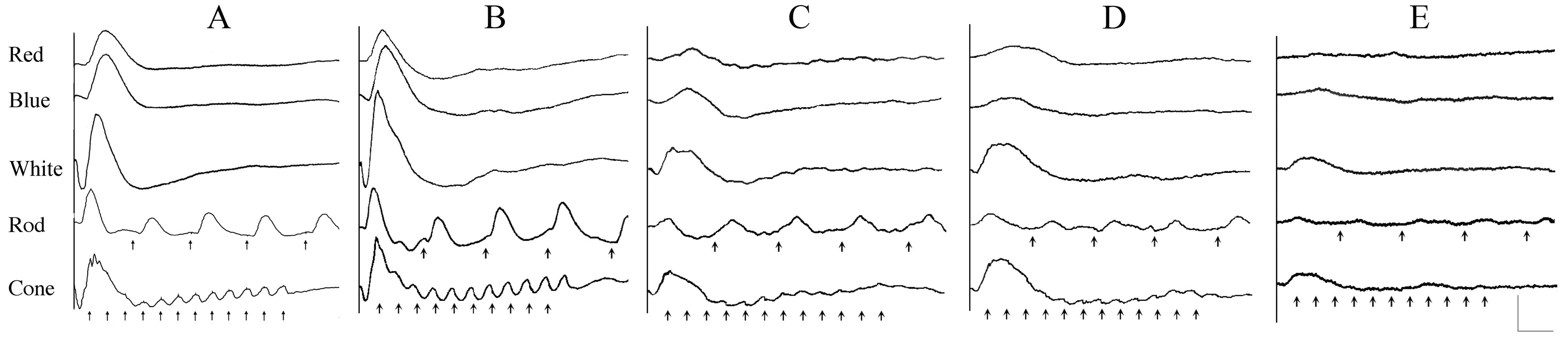
 Figure 1 of Goldstein, Mol Vis 2010; 16:1549-1569.