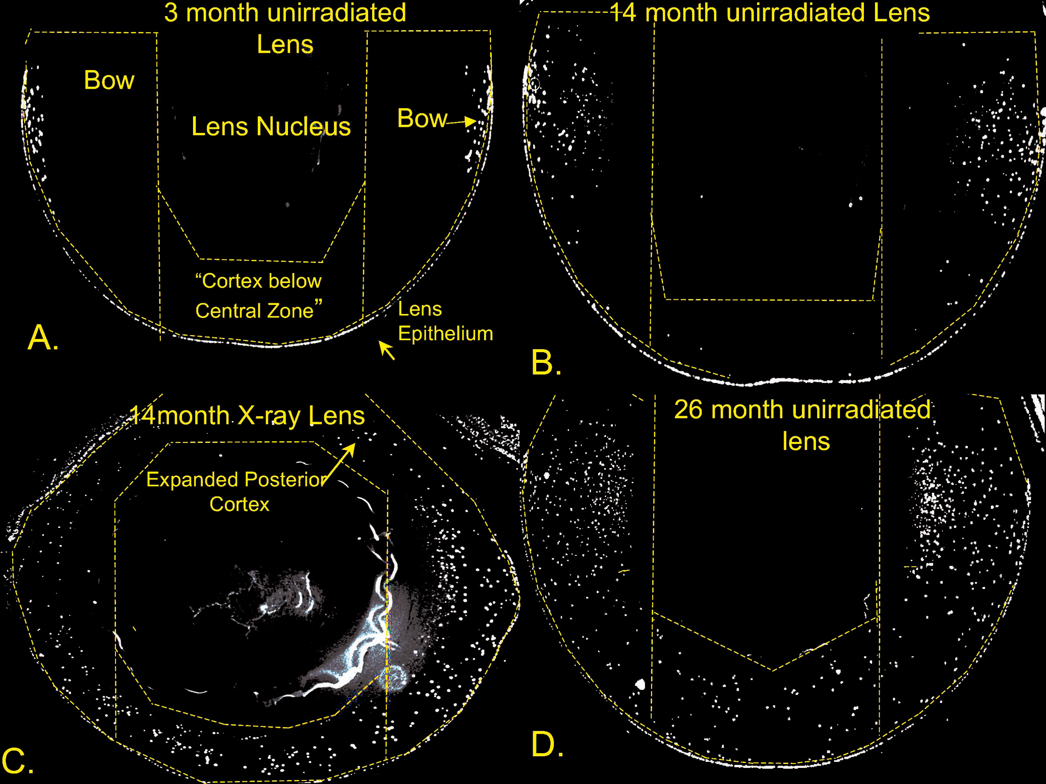
Figure 8. Typical images of DAPI-stained
paraformaldehyde-fixed sections from lenses unirradiated control lenses
and lenses from mice X-irradiated at 3-month of age. Panel
A was
from
unirradiated 3-month controls, panel
B was from 14 month
unirradiated controls, panel
C was from 14-month X-irradiated
lens, and panel
D was from a 26-month old unirradiated control.
Each image was derived from a panorama taken with a 10× objective (see
Methods). The dashed lines show the regions of the lenses used for
counting DNA fragments in different regions of cortex (see
Figure 9A,B).
These
include both bow regions, and the cortex below the central zone.
The posterior cortex was included if nuclear fragments were present.
The non-lens portions of the eye have been deleted for clarity.
Original magnification was 200×.
 Figure 8 of Pendergrass, Mol Vis 2010; 16:1496-1513.