Figure 7. A comparison of nuclei and
nuclear cortical fragments in a typical old-mouse eye section using
both DAPI and H&E stains. DAPI (A) and H&E (B)
staining are compared. All of the included nuclei stained with DAPI
(white) were also stained blue by Hematoxylin. All other H&E and
DAPI images demonstrated the same correspondence (not shown), but DAPI
was much easier to score at lower magnification so was generally used
in counting nuclei and nuclear fragments. Original magnification was
200×.
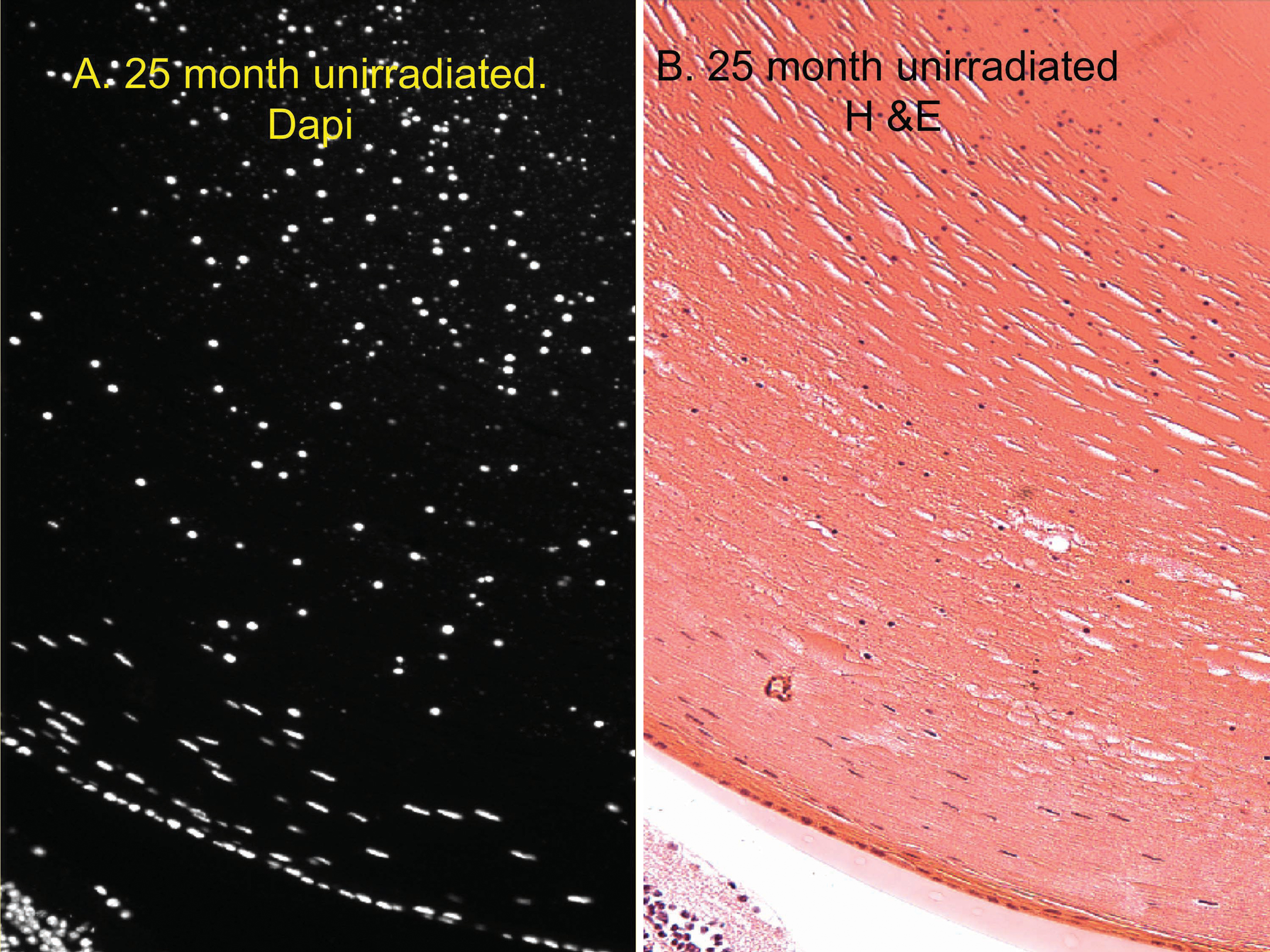
 Figure 7 of Pendergrass, Mol Vis 2010; 16:1496-1513.