Figure 5. Typical vital staining of the
anterior and posterior cortices of 10-month old mice lenses for DNA,
ROS, and cataract. DNA (Hoechst 33342) in the lenses is shown in red,
ROS (oxidized DHR) is shown in green, and cataract reflected light in
white. The LSCM (10× objective) was focused 120 microns below the
anterior poles of unirradiated control lenses (upper panel) and
X-irradiated lenses (lower panel) at the same age. The donors of the
lenses were 3 months old when irradiated and 10 months old when
analyzed. The first column depicts the DNA fluorescence in red (Hoechst
33342 fluorescence), middle Column shows ROS in green (oxidized DHR).
The right column shows light reflected from cataracts. Original
magnification was 200×.
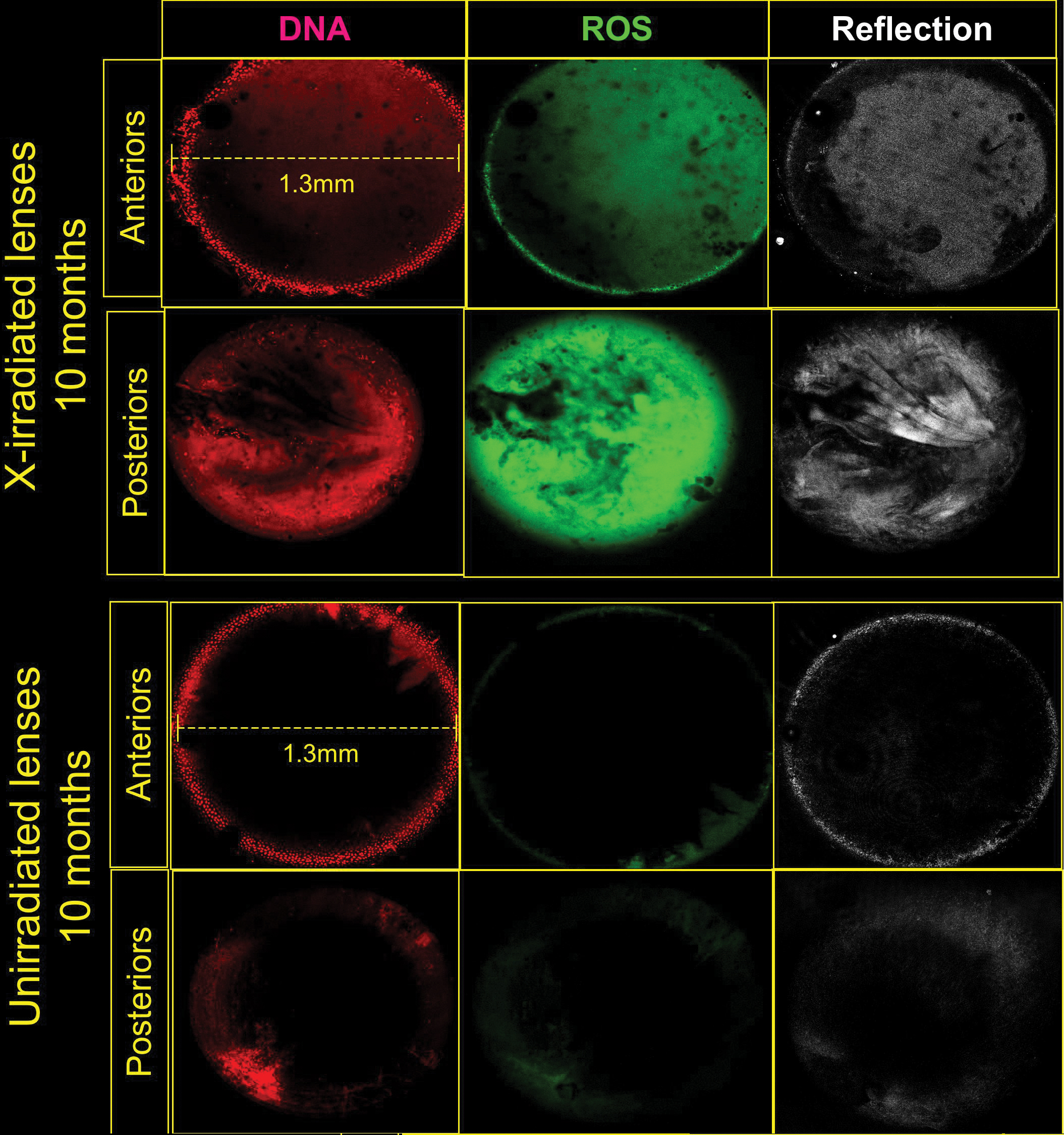
 Figure 5 of Pendergrass, Mol Vis 2010; 16:1496-1513.