Figure 3. Decreases in mouse surface LEC
cell density with age and X-irradiation using vital dye staining of
whole lenses. The cell density (cells/mm2) was determined by
counting nuclei stained with the vital dye Hoechst 33342 on the lens
surface in unirradiated controls (green squares) and X-irradiated mouse
lenses (red circles). X-Irradiation of these mice was at 3 months of
age. The error bars represent the standard error of the means, and
stars indicate a significant difference from age matched control values
using 1 tail Mann–Whitney test (the p values: **, p<0.02, ***,
p<0.002). Each point represents the mean of 4–6 mice.
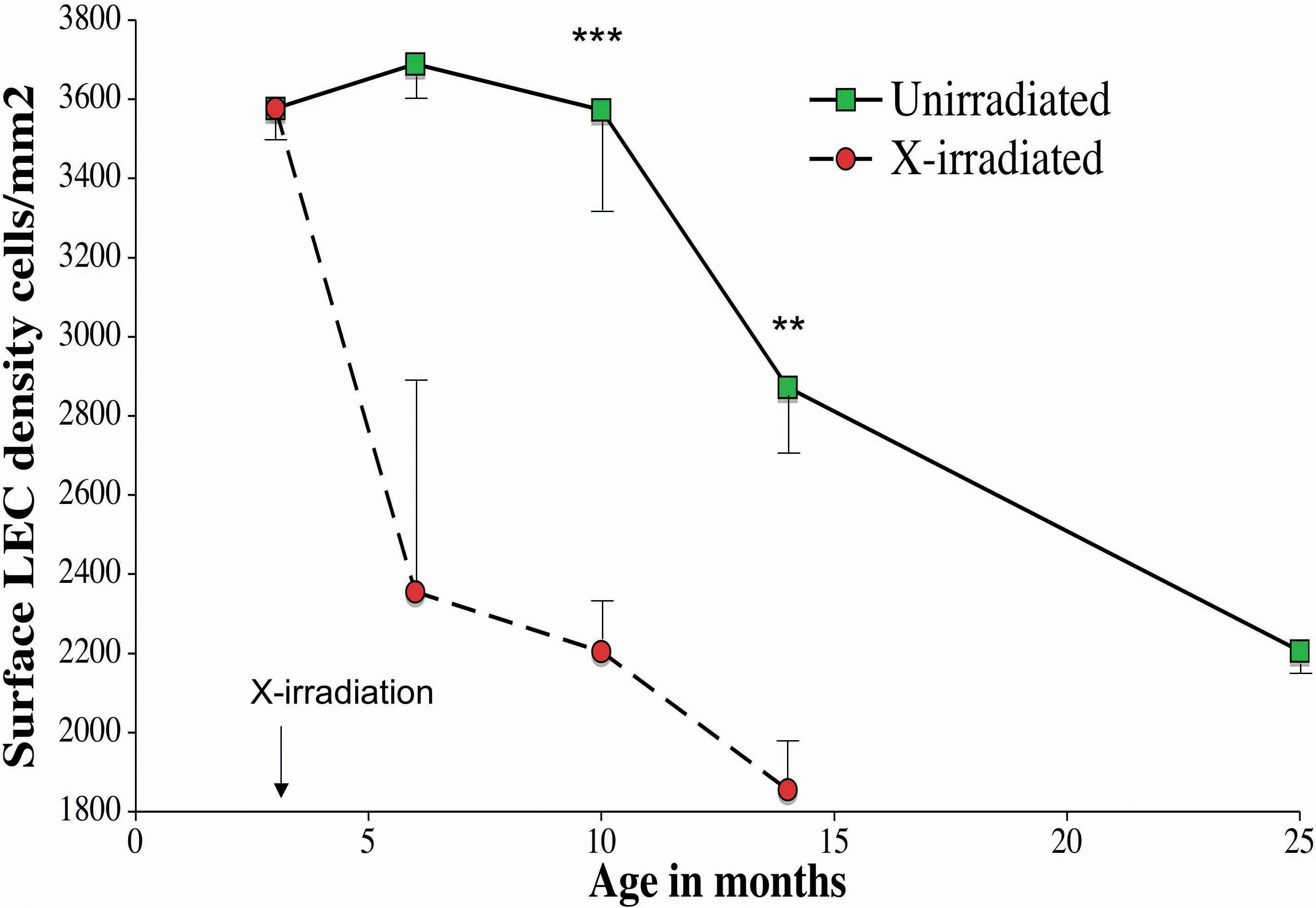
 Figure 3 of Pendergrass, Mol Vis 2010; 16:1496-1513.