Figure 12. The number of 8-OH-dG positive
cortical nuclear fragments found beneath the central zone. Fixed lens
sections were stained with antibodies to 8-OH-dG (green) and Dapi (Red)
as described in the methods (see also figure 11). The number of 8-OH-dG
positive nuclear fragments resistant to 2N HCl lying only in the cortex
below the Central Zone were counted and compared. The 8-OH-dG positive
fragments (green) increased with age and were significantly higher in
the X-irradiated lenses at 12.5 months and 14 months. Five to 7 of
samples were analyzed at each age, except the 3-month X-ray with 2 mice
lenses; and the 7-month X-ray with 4.
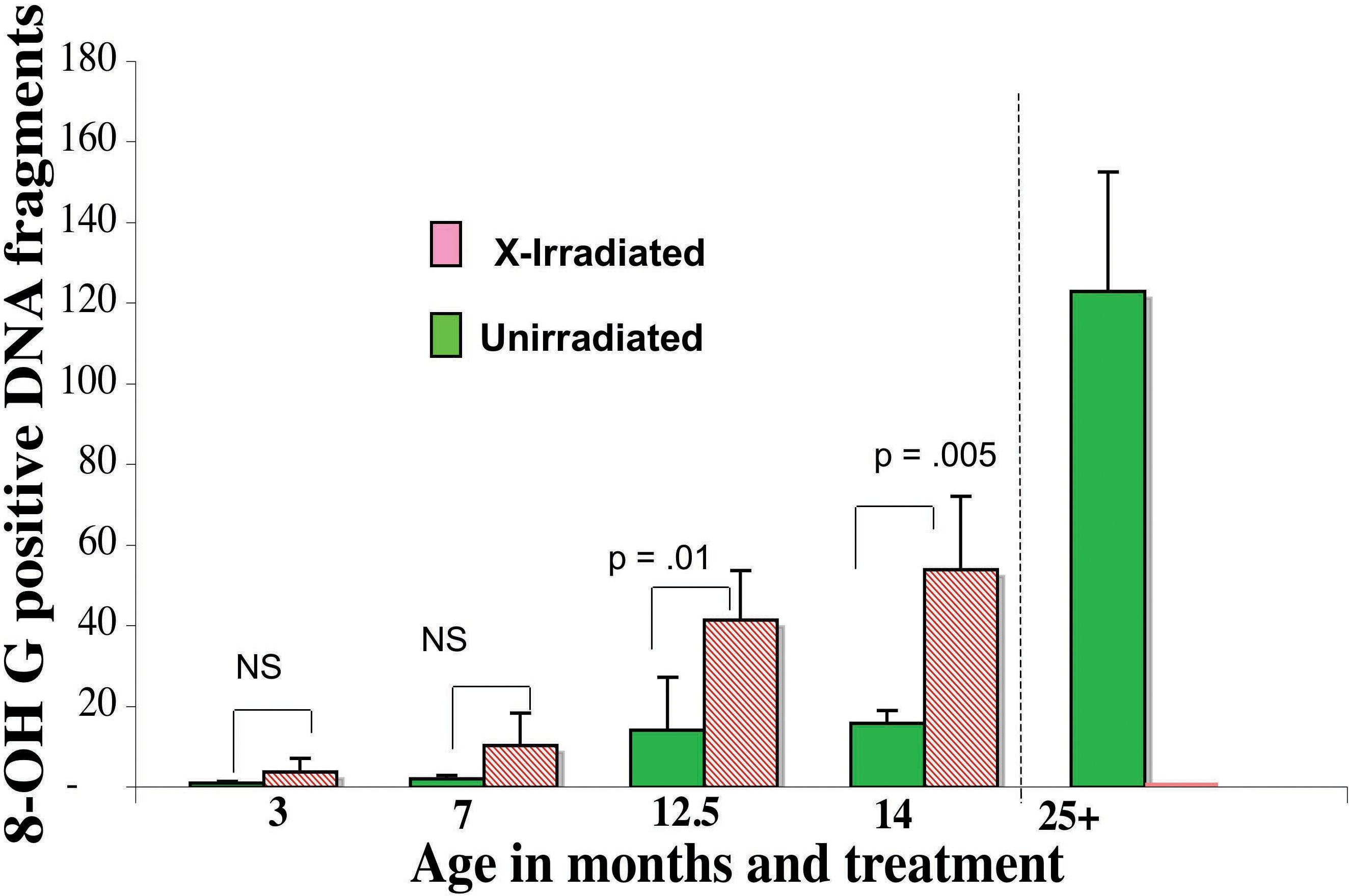
 Figure 12 of Pendergrass, Mol Vis 2010; 16:1496-1513.