Figure 10. Fixed-eye mouse lens sections
were stained with; antibodies to alpha smooth muscle actin shown in
green, and aquaporin 0 shown in red and also DNA shown in blue as
described in the Methods. Typical staining of lens fibers with
aquaporin O antibodies (red) and myofibroblast-like cells (green) are
shown. Note that aquaporin O comprises approximately 60% of lens fiber
membrane proteins, and that myofibroblasts are normally not present in
the normal young mouse lens (see text). A: Posterior of
14-month X-irradiated lens, arrows point out myofibroblasts (green). B:
Posterior
region of typical old (26-month) mouse lens lacking
myofibroblasts. Panels C-G show images of the bow
regions of irradiated or unirradiated control lenses of various ages
that were all negative for myofibroblasts. Original magnification was
200×.
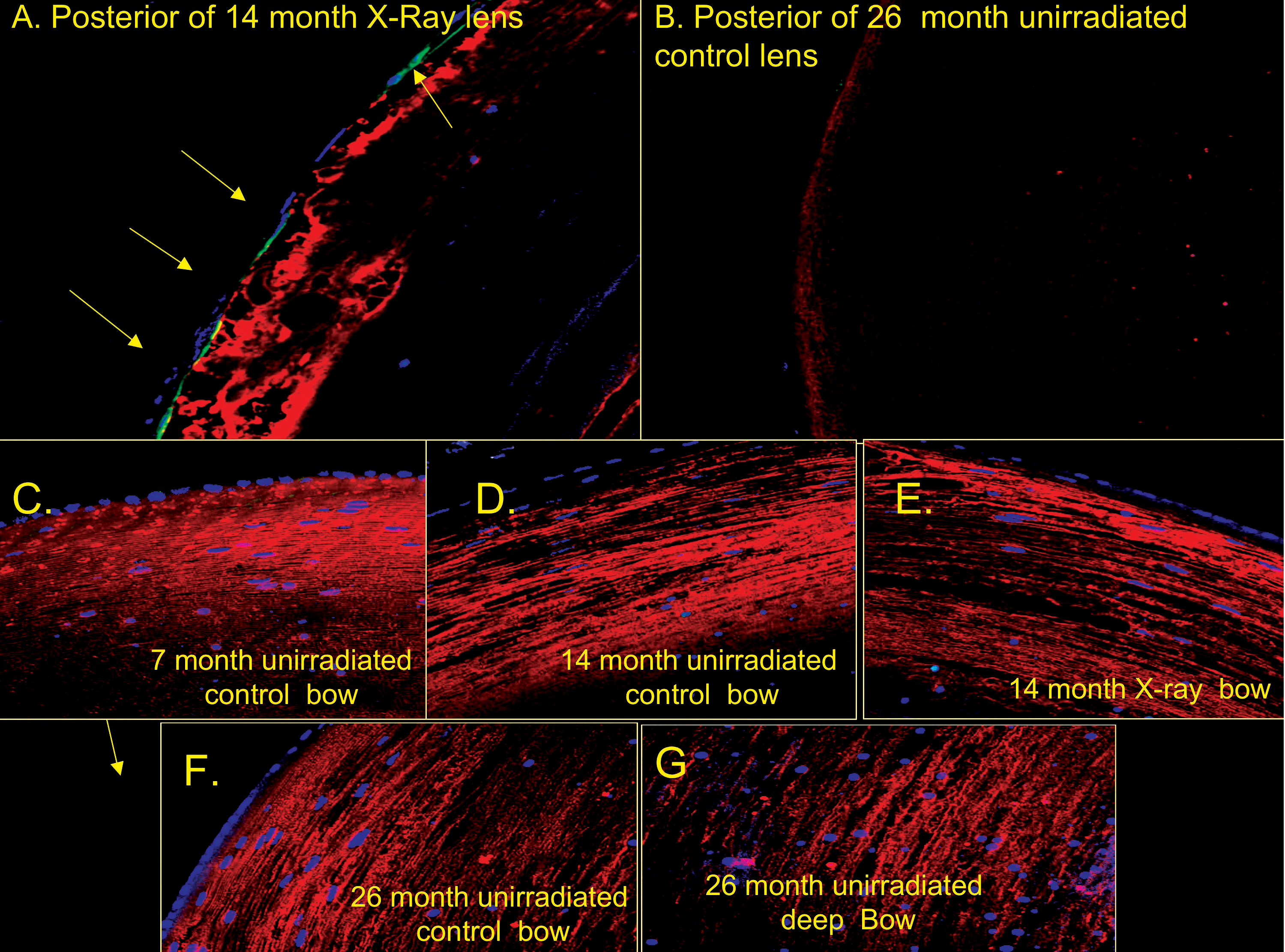
 Figure 10 of Pendergrass, Mol Vis 2010; 16:1496-1513.