Figure 1. Resveratrol protects against H2O2-induced cell death and apoptosis in HLEB-3 cells. A: The HLEB-3 cells were incubated with different concentrations of RES (2.5, 5.0, 10.0, and 20.0 μM) for12 h before H2O2 (100 μM) treatment for 24 h. B: The cells were incubated with 20.0 μM RES for different time (12, 24, 48 h), then treated with 100 μM H2O2 for 24 h. C: The cells were pretreated with 20.0 μM RES for 12 h,then exposed to different concentration of H2O2 (50, 100, 200 μM) for 24 h, the cell viability was measured by WST-1 assay. The data are represented as means±SD from three
independent experiments. The asterisk indicates that p<0.05 compared to the untreated control. D: The cells were incubated with 10.0 and 20.0 μM RES for 12 h then treated with 100 μM H2O2 for 24 h. Flow cytometric analysis was used to quantify the rate of cell apoptosis using double staining of Annexin V-FITC
and PI. The result is one representative example of three separate experiments. E: Resveratrol (20μM) significantly decreased the apoptosis rate of HLEB-3 cells, **p<0.01 versus positive control (t-test), the preventative effect of 20 μM RES was improved versus 10 μM RES,*p<0.05.
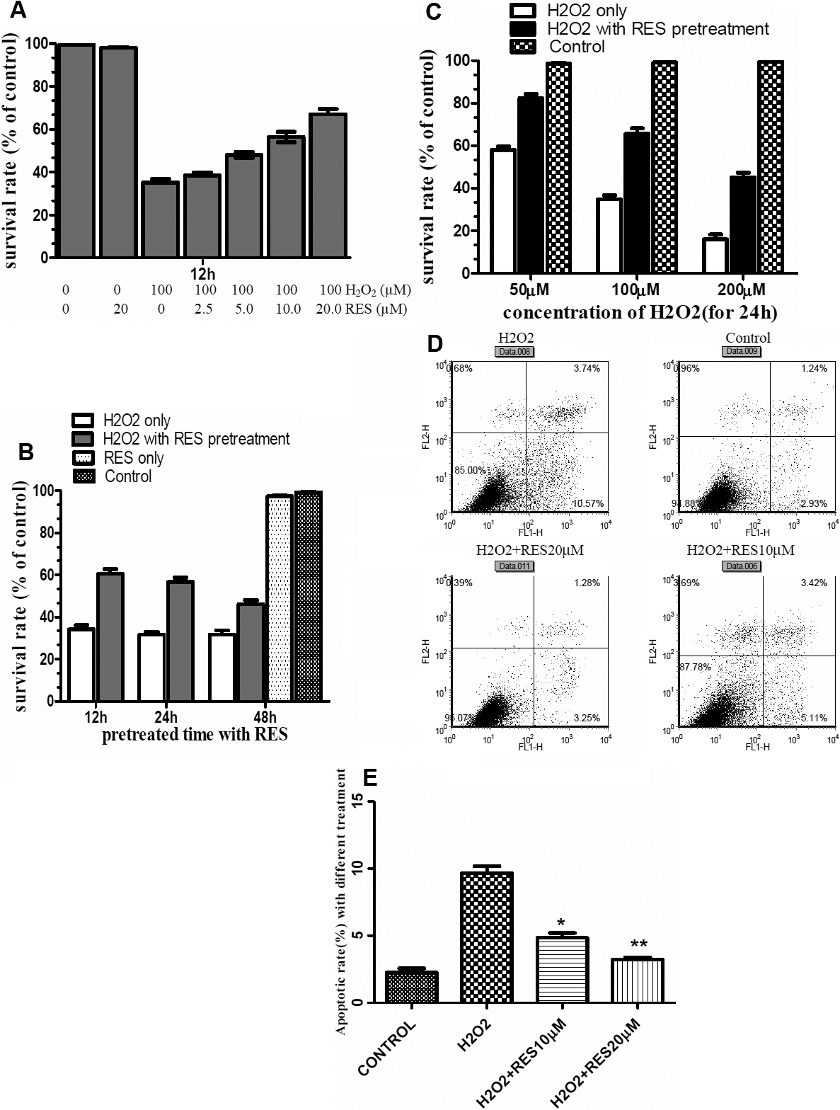
 Figure 1 of
Zheng, Mol Vis 2010; 16:1467-1474.
Figure 1 of
Zheng, Mol Vis 2010; 16:1467-1474.