Figure 3. Vascular endothelial growth
factor concentrations in the vitreous body and serum from proliferative
diabetic retinopathy and macular hole patients. The VEGF concentrations
in the vitreous were much higher in PDR than in MH patients. In
contrast, the VEGF concentrations in the serum were not significantly
different between PDR and MH patients. **, p<0.01 versus MH
(vitreous body; Tukey test).
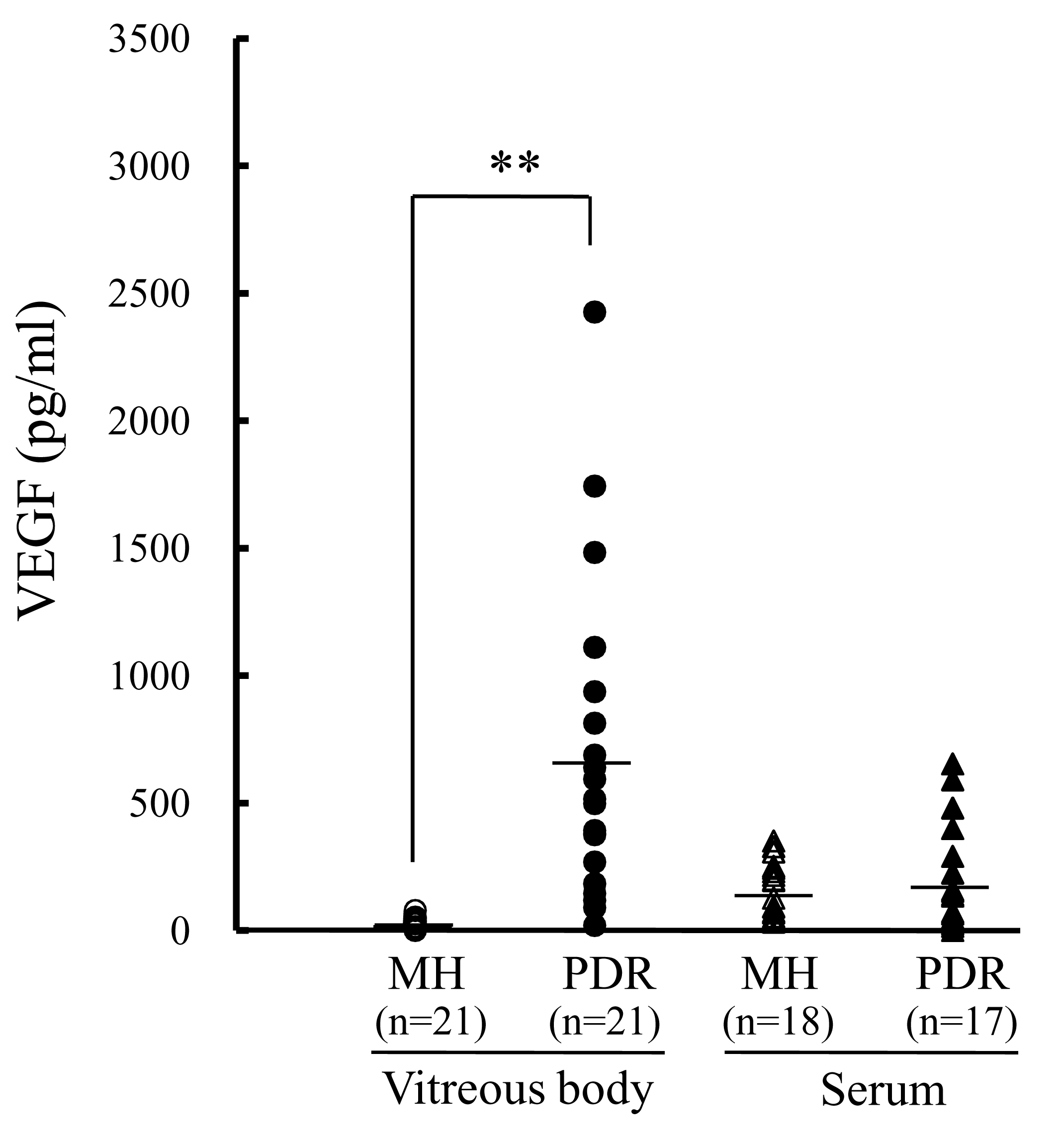
 Figure 3 of Izuta, Mol Vis 2010; 16:130-136.