Figure 9. Schematic model depicting the rapid shift in migration patterns of posterior fiber ends during the initial stages of PSC formation
in RCS/Lav rat lenses. A, D: Initially, the posterior suture pattern (gray) is a three branched inverted Y (A), wherein fiber ends (D; gradations of purple) migrate either along meridians to the proximal ends of the suture branch or along a curved path to
the lateral aspect of the suture branch. Successive cohorts of migrating fiber ends are depicted in decreasing intensities
of color, with the oldest generation denoted by the darkest color. B, E: The first noticeable change is the rapid formation of abnormal suture sub-branches (B), due to the change in migration paths of selected groups of fiber ends (E). C, F: Over time, the elaboration of abnormal sub-branches continues (F) as migration paths undergo additional shifts through successive cohorts of fiber ends.
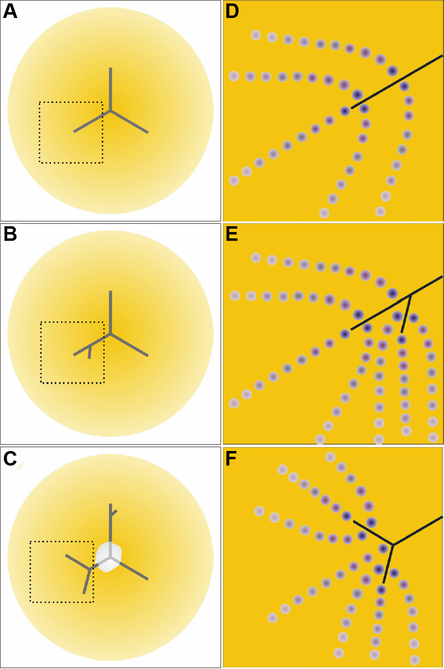
 Figure 9 of
Joy, Mol Vis 2010; 16:1453-1466.
Figure 9 of
Joy, Mol Vis 2010; 16:1453-1466.