Figure 8. Schematic representation of the posterior fiber ends of RCS/Lav rat lenses depicting the change in fiber end morphology, filopodial
configuration and F-actin distribution as a function of time. A-C: Note the change in fiber end shape from irregular spheroids having short, unidirectional filopodia at 2-3 weeks of age to
enlarged, disorganized ends with elongated filopodia at 4–6 weeks. By 7–8 weeks, the ends were extremely dilated and globular
with complete absence of filopodia. The concomitant F-actin rearrangements at the corresponding ages are also diagrammatically
summarized in panels D, E, and F. At 2–3 weeks of age, F-actin distribution was predominantly peripheral within the BMC (D), however by 4–6 weeks, foci were common (E) within the forming PSC plaque. The F-actin was entirely rearranged into rosettes within the dilated fiber ends (F).
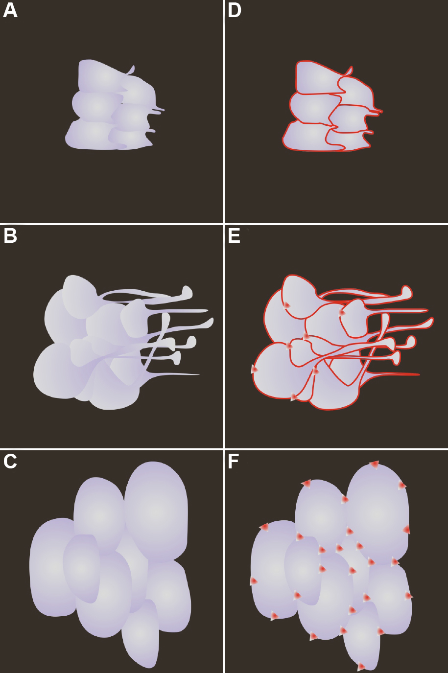
 Figure 8 of
Joy, Mol Vis 2010; 16:1453-1466.
Figure 8 of
Joy, Mol Vis 2010; 16:1453-1466.