Figure 5. Scanning electron micrographs of RCS/Lav lenses showing the morphology and arrangement of the posterior fiber ends (7–8 weeks).
A, B: Low and high magnification view of the PSC plaque. The arrow points to the original suture plane which is eventually obscured
by the PSC, as a result of a cessation of fiber ends migration. The ends within the plaque are dilated/globular with loss
of end to end interaction. C, D: Some PSC plaques maintained the end to end interaction, but were severely dilated and had completely lost their filopodia.
Panel D also clearly depicts the variability in fiber end size that is seen at this age. E, F: Some areas which were more severely affected showed stellate shaped ends with surrounding areas of apparent fiber end loss
(panel F-asterisks).
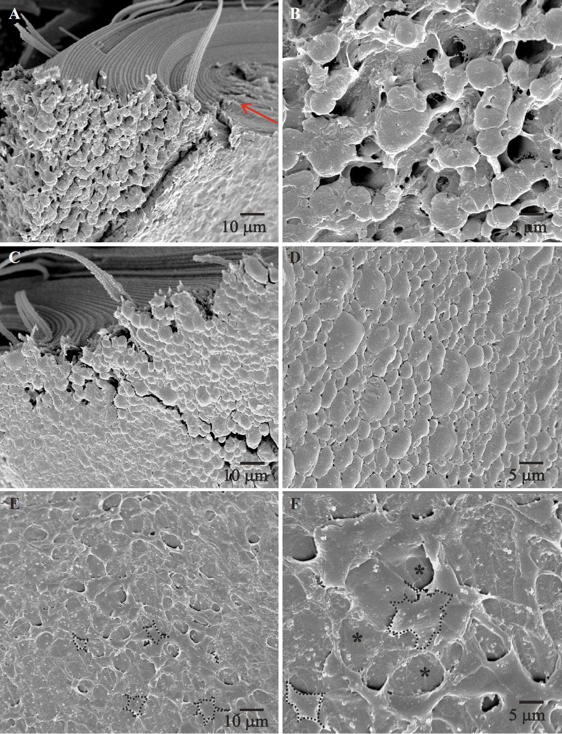
 Figure 5 of
Joy, Mol Vis 2010; 16:1453-1466.
Figure 5 of
Joy, Mol Vis 2010; 16:1453-1466.