Figure 4. Scanning electron micrographs of RCS/Lav lenses showing the morphology and arrangement of the posterior fiber ends (2–6 weeks).
A, B: Low and high magnification images showing the normal shape and arrangement of the basal ends at 2 weeks of age. Note the
uniform size and orientation of the filopodia in panel B (arrows). C, D: Low and high magnification images at 3 weeks postnatal, showing some areas with domed ends (asterisks in panel D). For the most part, the ends continue to maintain their normal shape and arrangement as seen at 2 weeks of age. E, F: By 4 weeks postnatal, fiber ends show distinct changes in their shape, end to end adhesion and the filopodia. Panel E shows the border between ends within the forming opacity (left of the dotted line) and normal ends just outside the opacity.
The disordered and lengthened filopodia, membrane blebbing and loss of end to end adhesion can be clearly seen in Panel F.
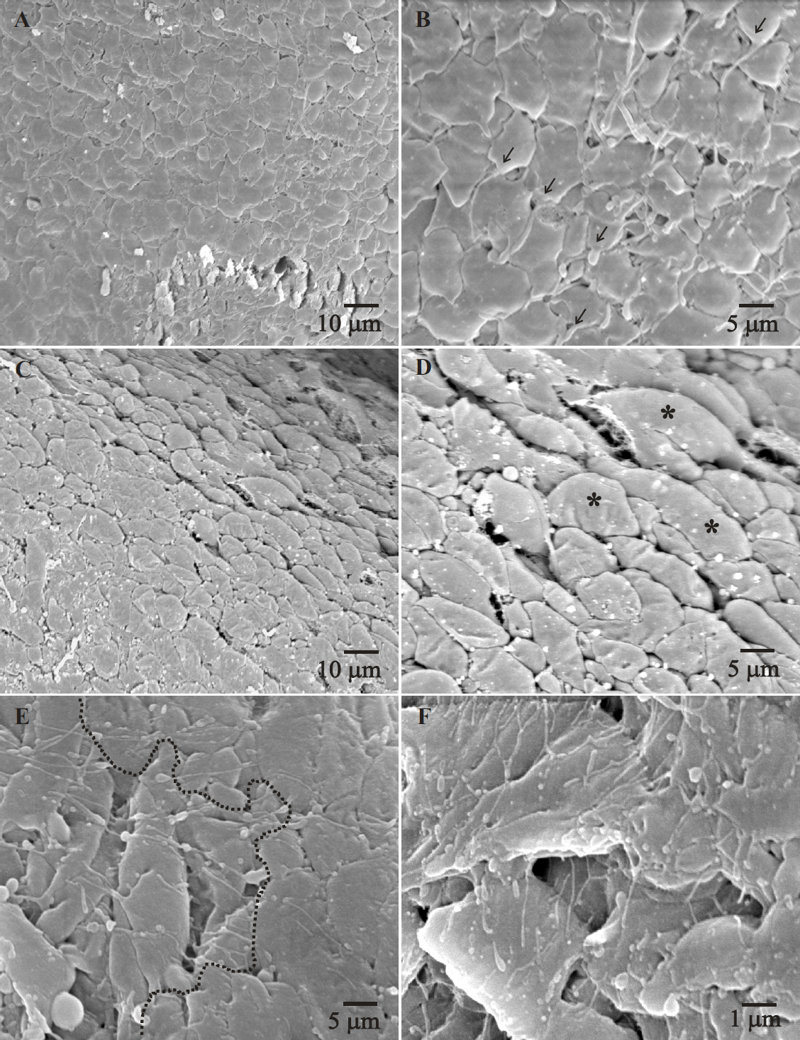
 Figure 4 of
Joy, Mol Vis 2010; 16:1453-1466.
Figure 4 of
Joy, Mol Vis 2010; 16:1453-1466.