Figure 2. Dissecting microscope photographs of unfixed dystrophic (A-F) and control (G-I) RCS rat lenses. A: Prior to PSC development, lenses from 2 week old animals displayed three-branched inverted-Y sutures on the posterior surface.
B: By 4 weeks, one or more abnormal suture sub-branches was apparent (arrows). C, D: Opacities which circumscribed the suture branches were detected as early as 5 weeks post natal. E: The opacity continued to grow through 6 weeks of age. F: The PSC completely obscured the suture by 7–8 weeks postnatal. In contrast, the control lenses showed no change from the
normal inverted Y suture with age (G: 2–3 weeks, H: 4–6 weeks, I: 7–8 weeks).
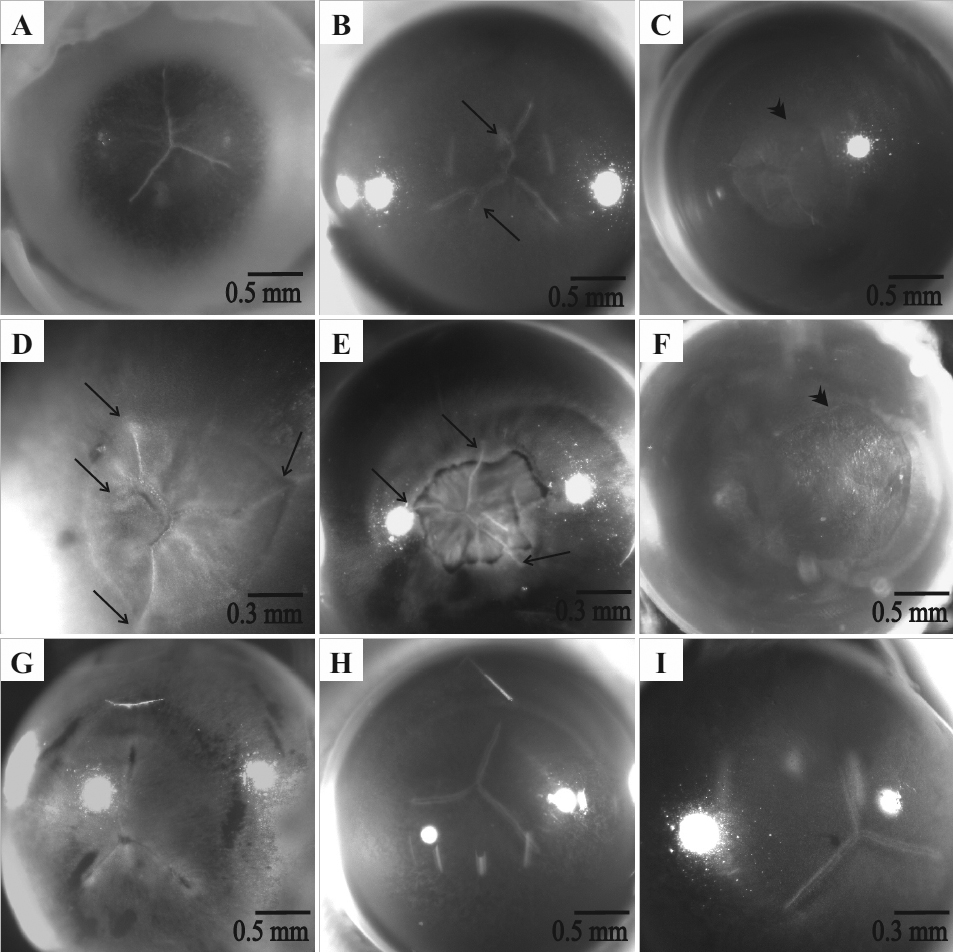
 Figure 2 of
Joy, Mol Vis 2010; 16:1453-1466.
Figure 2 of
Joy, Mol Vis 2010; 16:1453-1466.