Figure 3. Dose–response effect of NGF and ANA on VEGF expression in culture medium and cell pellet of HCECs after supplementation of
1, 10, and 100 ng/ml of NGF or ANA at 1, 10 and 100 μg/ml of medium. NGF enhances the presence of VEGF both in the medium
and in pellet at concentration of 1 or 10 ng/ml of NGF. At 100 ng/ml NGF has an inhibitor action on VEGF expression. ANA exposure
has no effect on the concentration of VEGF in the medium (C), but significantly reduces (*p<0.05) VEGF presence in the pellet at concentration of 100 ug/ml (D). Confocal microscopic analysis of cells exposed to 10 ng/ml of NGF and 100 µg/ml of ANA are illustrated, respectively in
F and G. Note the enhanced expression of VEGF after exposure of NGF (F) and the down-regulation after exposure of ANA (G), compared to control (E). Arrows point to VEGF immunopositivity. Scale bars: E-G=15μm.
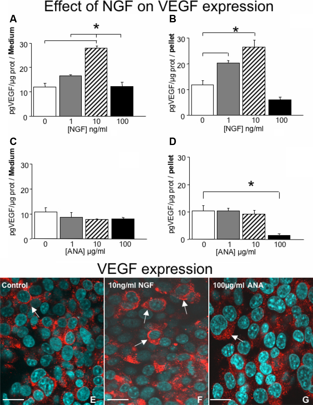
 Figure 3 of
Sornelli, Mol Vis 2010; 16:1439-1447.
Figure 3 of
Sornelli, Mol Vis 2010; 16:1439-1447.