Figure 3. : Protein modeling studies
showing the superimposition image of L559V mutant (yellow) and wild
type conformers (cyan). Only the mutant structures in which deviations
were observed are shown. The conformational changes in the secondary
structure elements are marked for the amino acid residues 544–549 (A),
552–560 (B) and 563–572 (C), respectively. The changes were mainly
observed in non-covalent hydrophobic interactions.
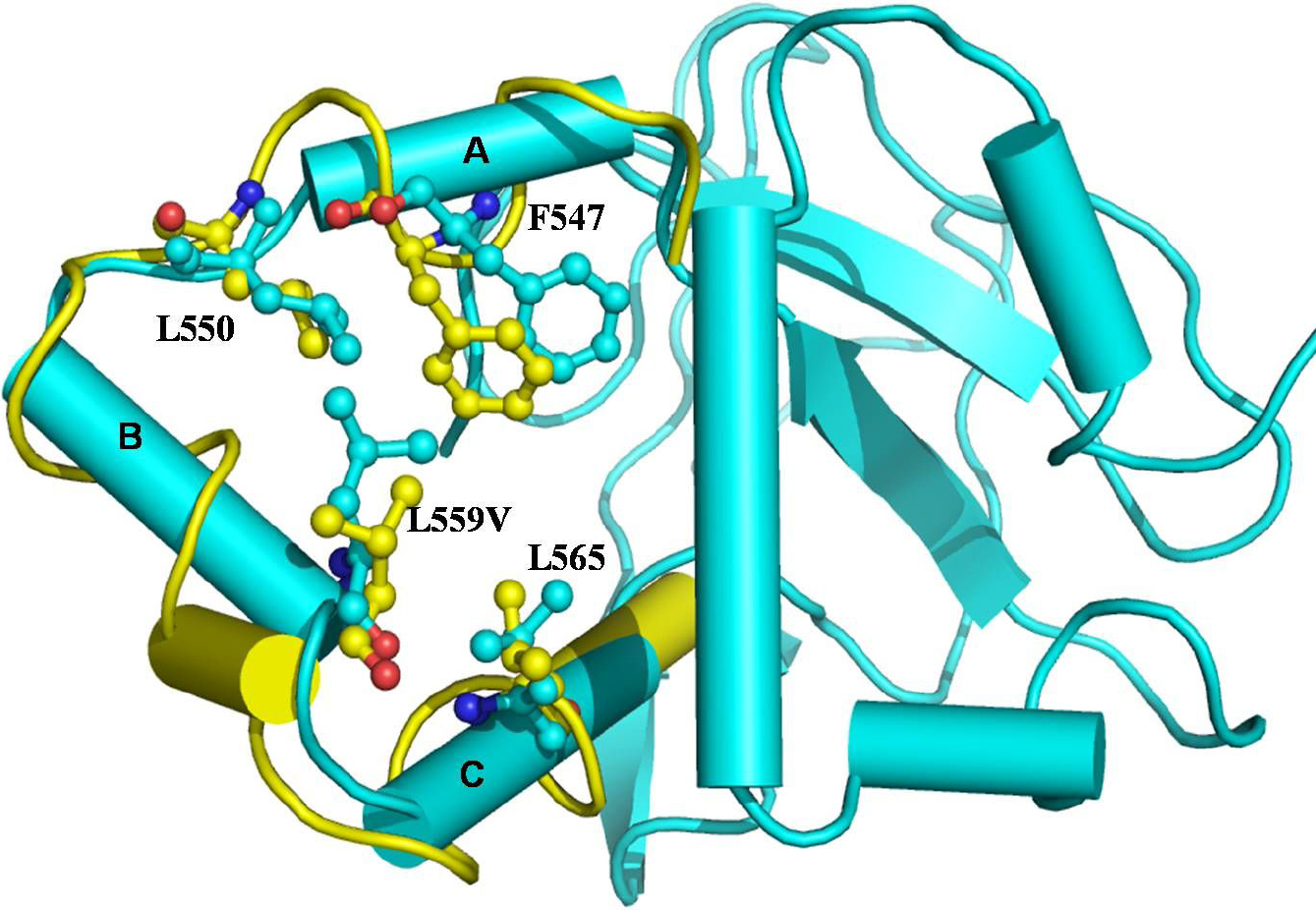
 Figure 3 of Paliwal, Mol Vis 2010; 16:1429-1438.