Figure 2. Family 2 showing the L559V
mutation in the transforming growth factor beta induced (TGFBI)
gene. A: Pedigree of the family. Filled boxes represent
affected individuals. Open boxes represent unaffected individuals.
Arrowhead indicates the proband. Gray boxes mark the affected
individuals with a variable phenotype. B-E: Slit lamp
and in-vivo confocal photomicrographs of the affected individual. The
representative clinical photographs of the proband show the presence of
a map-like structure in the right eye (B). Retro illumination
photomicrograph demonstrates a fingerprint-like pattern in the right
eye (C). The left eye of the proband shows multiple dot-like opacities
(D). The map pattern in the sub-epithelial region, which was confirmed
by in vivo confocal microscopy, is shown (2E). F: Partial
nucleotide sequence of the transforming growth factor beta induced (TGFBI)
gene.
The chromatogram of the patient (P) is shown compared to a
control (C). A heterozygous T>G substitution, marked by the
arrowhead, is shown. The block denotes the nucleotide with missense
mutation that results in amino acid substitution of Leucine at 559
position with Valine. The partially dashed blue line on the top right
of the chromatogram marks the start of intron while the red line marks
the exonic region. G-H: Representative slit lamp
photomicrographs of the affected family member. Representative clinical
photographs of the affected family member (G) showing very fine
dot like opacities in the right eye. The indirect slit lamp
retroillumination image (H) shows the presence of multiple
dimple-like structures in the affected cornea.
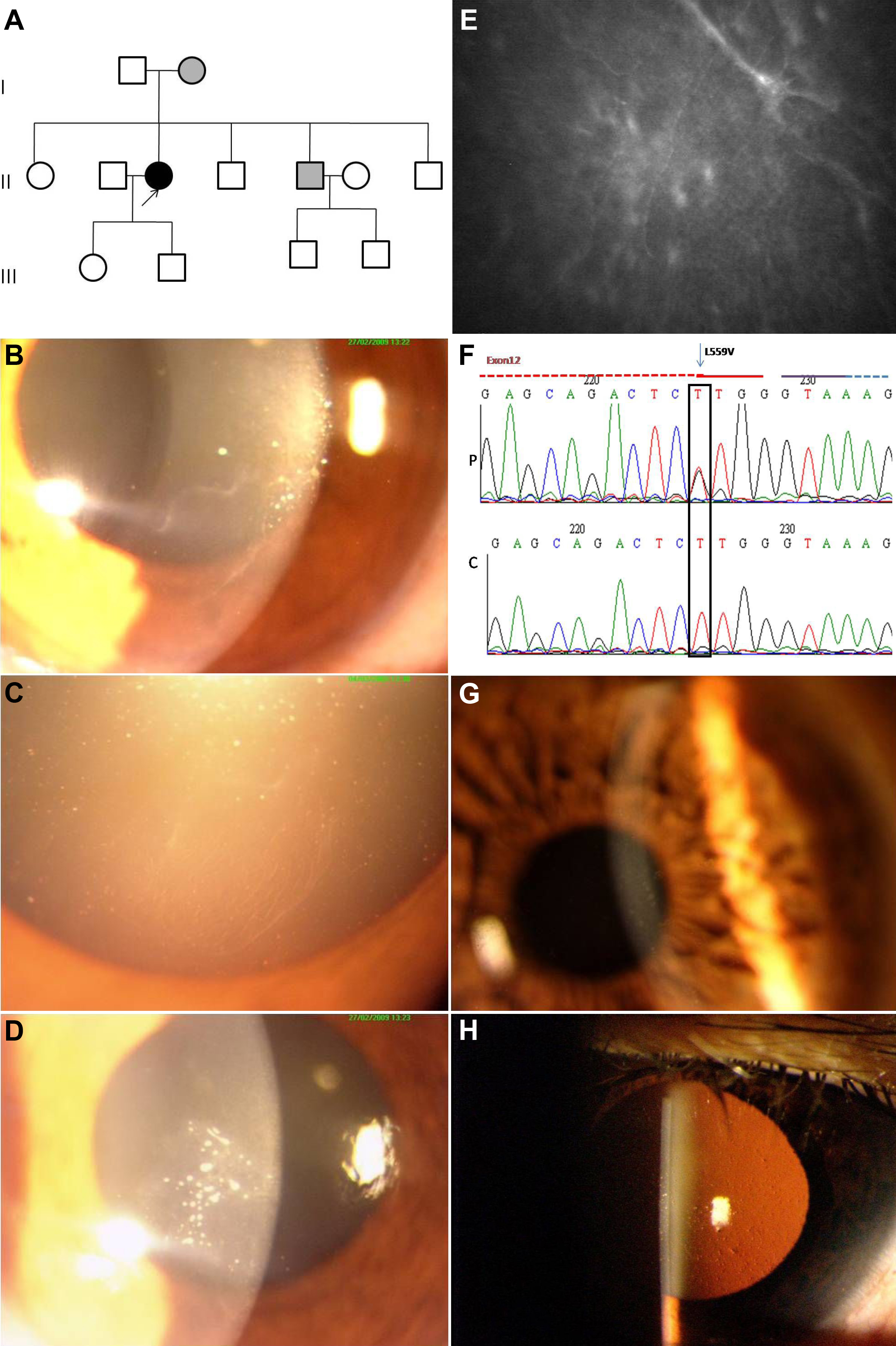
 Figure 2 of Paliwal, Mol Vis 2010; 16:1429-1438.