Figure 1. Family 1, showing the S516R
mutation in the transforming growth factor beta Induced (TGFBI)
gene. A: Pedigree of the family. Filled boxes represent
affected individuals. Open boxes represent unaffected individuals.
Arrowhead indicates the proband. Filled circle with a slash indicates a
deceased individual. B: Slit lamp photomicrograph of the
affected individual. The representative clinical photograph shows the
presence of discrete gray-white deposits in the right eye of the
proband, with clear intervening stroma resembling granular corneal
dystrophy. C: Partial nucleotide sequence of the transforming
growth factor beta induced (TGFBI) gene. The chromatogram of the
patient (P) is shown compared to a control (C). A heterozygous C>G
substitution, marked by the arrowhead, is shown. The black box denotes
the nucleotide that causes the missense mutation resulting in amino
acid substitution of the Serine (S) at amino acid position 516 with
Arginine (R). The partially dashed blue line at the top left of the
chromatogram marks the intronic region, while the red line on the right
marks the start of the exon. D: Protein modeling in S516R
mutation showing the superimposition of S516R mutant (yellow) and wild
type conformers (cyan). Only the mutant structures, where the
deviations were observed, are shown in the figure. The conformational
changes in the secondary structure elements are shown for amino acid
residues 505–511 (A), 516–525 (B) and 544–550 (C). The changes observed
in molecular interactions (Hydrogen bonds) are also marked by dashed
lines.
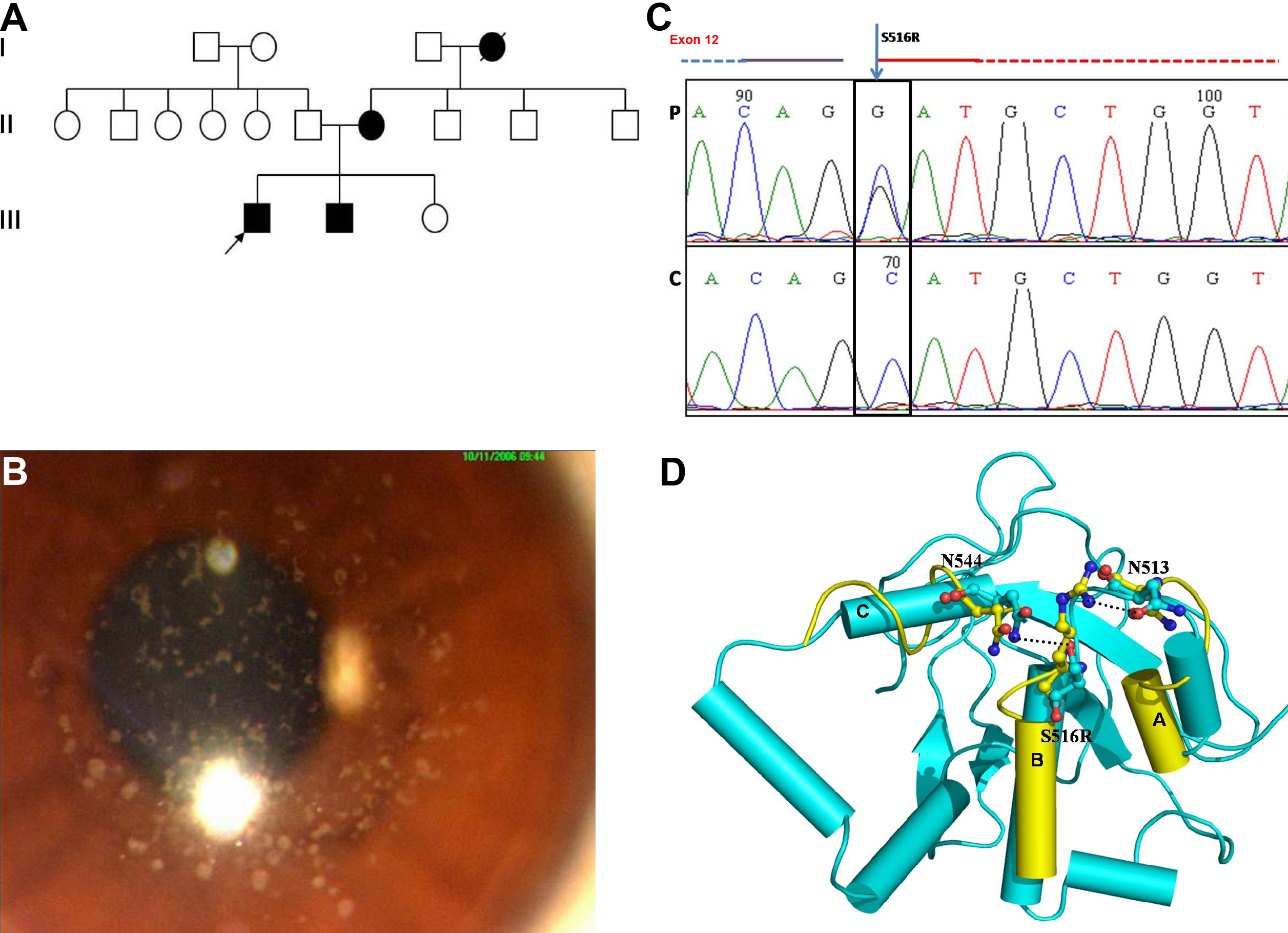
 Figure 1 of Paliwal, Mol Vis 2010; 16:1429-1438.