Figure 7. Inner limiting membranes of
protein O-mannose N-acetylglucosaminyltransferase 1 (POMGnT1) knockout
and Largemyd mice exhibit reduced elasticity. Flatmounted
inner limiting membranes from postnatal day 2 (P2) newborn mice were
immunostained with anti-laminin. AFM indentation experiments were
performed to determine the Young’s modulus. Micrographs of laminin
immunofluorescence staining of wild type (A), protein O-mannose
N-acetylglucosaminyltransferase 1 knockout (B) and Largemyd
mice (C) are shown. Note that the mutant inner limiting
membranes showed many holes (B and C). D and E
are representative force displacement curves for indentation
experiments on the inner limiting membranes of protein O-mannose
N-acetylglucosaminyltransferase 1 knockout and Largemyd
mice. Apparent Young’s modulus measurements from inner limiting
membranes isolated from 4 mice for each genotype are shown in F.
The
bars represent means with standard deviations (p<0.01, ANOVA).
When compared to the controls, significantly reduced Young’s modulus
was observed for both POMGnT1 knockout (p<0.01) and Largemyd
(p<0.01) inner limiting membranes (post hoc Student’s t-test).
Asterisks
in F indicate p<0.01. These results indicated that
the inner limiting membranes of protein O-mannose
N-acetylglucosaminyltransferase 1 knockout and Largemyd mice
exhibited reduced elasticity compared to the wild type. MPa represents
megapascal. nN represents nanonewton. The scale bar in C is
equal to 25 μm.
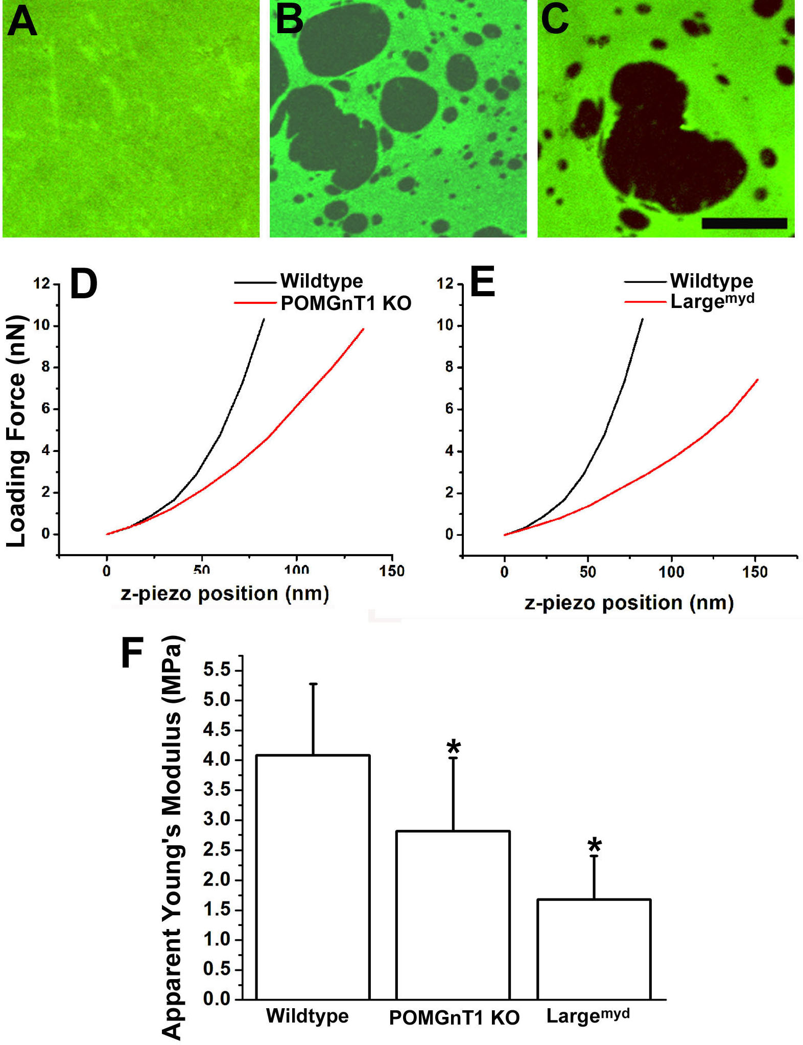
 Figure 7 of Hu, Mol Vis 2010; 16:1415-1428.