Figure 5. Inner limiting membrane
disruptions during early eye development in protein O-mannose
N-acetylglucosaminyltransferase 1 (POMGnT1) knockout mice. Electron
microscopic analysis was performed on the developing retinas at
embryonic day 11.5 (A and B), embryonic day 13.5 (C
and D), and embryonic day 15.5 (E and F). A,
C, and E are from wildtype retinas. B, D,
and
F are from knockout retinas. Note the broken inner limiting
membrane at E13.5 (arrow in D) and the absence of the inner limiting
membrane at E15.5 (asterisks in F) in POMGnT1 knockout mouse
retina. The scale bar in D is equal to 500 nm. +/+ represents
wild type; −/− represents homozygous knockout.
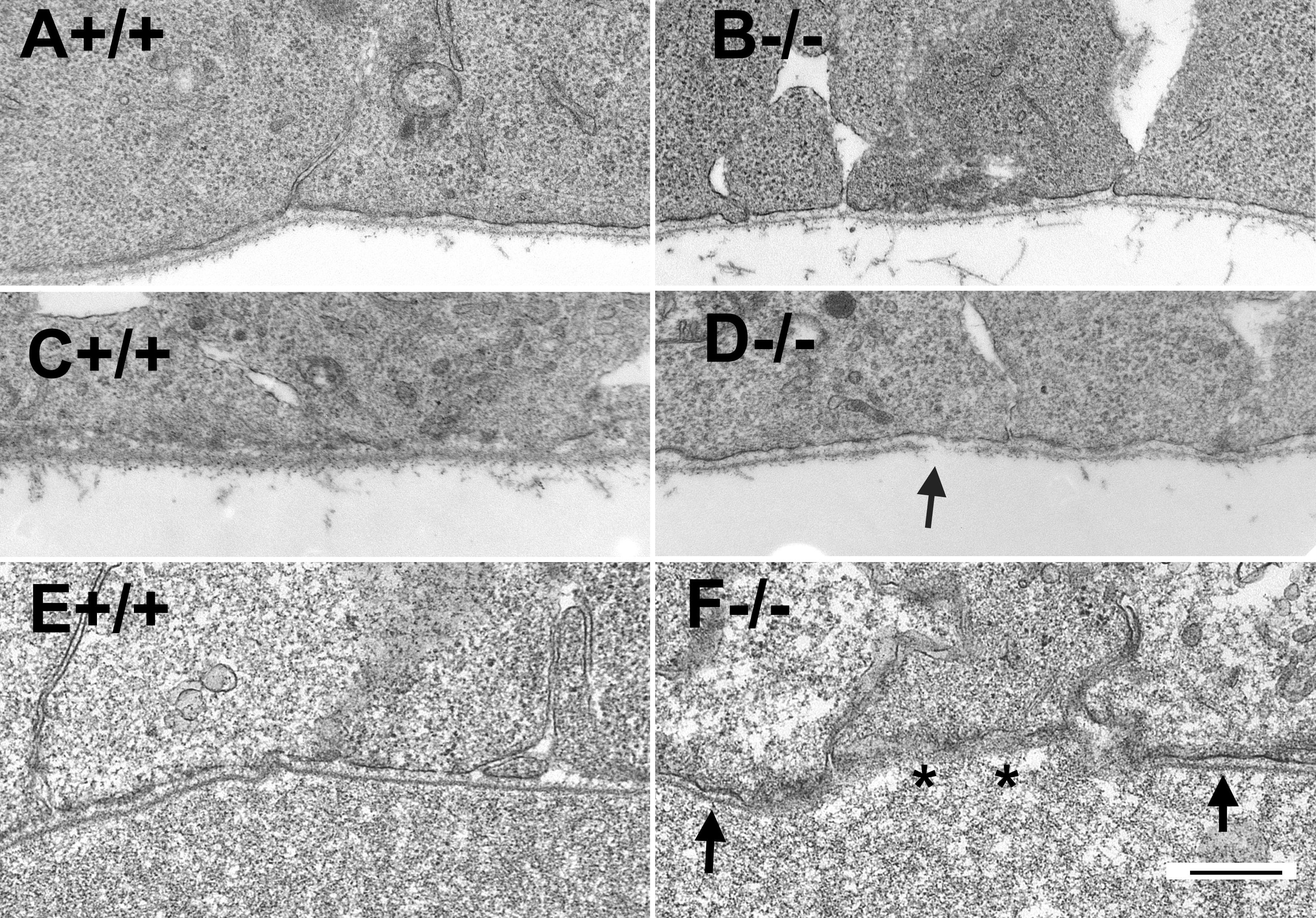
 Figure 5 of Hu, Mol Vis 2010; 16:1415-1428.