Figure 4. Morphological changes were
observed in the Müller glia of protein O-mannose
N-acetylglucosaminyltransferase 1 knockout retinas. Adult retinal
sections were immunostained with antibodies against cellular
retinaldehyde-binding protein (CRALBP; A and B), Kir4.1
(C and D), and aquaporin-4 (AQP-4; E and F).
Fluorescence
micrographs shown in A, C, and E
are from wild type retinas and fluorescence micrographs shown in B,
D, and F are from knockout retinas. Note the protrusion
of Müller glia endfeet intro the vitreous (arrows in B) and
reduced levels of Kir4.1 and AQP-4 at sites lacking the inner limiting
membrane (asterisks in D and F). The scale bar in F
is equal to 100 μm. Abbreviations: AQP-4 represents aquaporin-4; CRALBP
represents cellular retinaldehyde-binding protein; DAPI represents
4',6-diamidino-2-phenylindole; INL represent inner nuclear layer; ILM
represents inner limiting membrane; IPL represents inner plexiform
layer; ONL represents outer nuclear layer; OPL represents outer
plexiform layer.
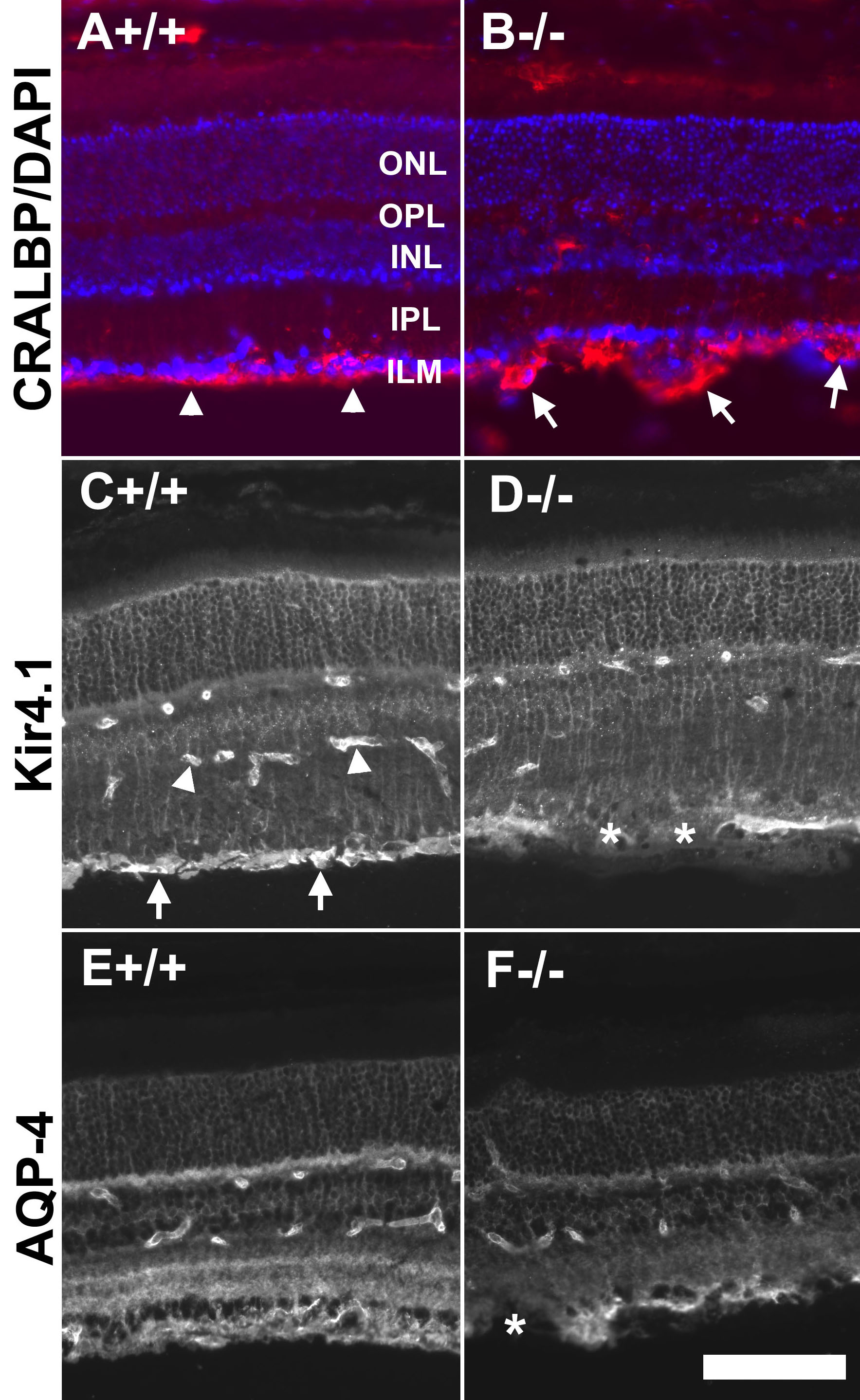
 Figure 4 of Hu, Mol Vis 2010; 16:1415-1428.