Figure 2. Disruptions of the inner
limiting membrane in protein O-mannose N-acetylglucosaminyltransferase
1 (POMGnT1) knockout retina. Transmission electron microscopy was
carried for the adult retinas. Micrographs of transmission electron
microscopy of wild type (A, C) and knockout (B, D)
retinas
are shown. Note that the wild-type inner limiting membrane was
continuous (arrowheads in A). Knockout inner limiting membrane
was discontinuous (arrows in B) with protrusion of Müller glial
processes into the vitreous humor. The knockout Bruch’s membrane
(arrowheads in D) was normal. The scale bar in B is
equal to 500 nm for A and B. The scale bar in D
is equal to 500 nm for C and D. +/+ represents wild
type; −/− represents homozygous knockout.
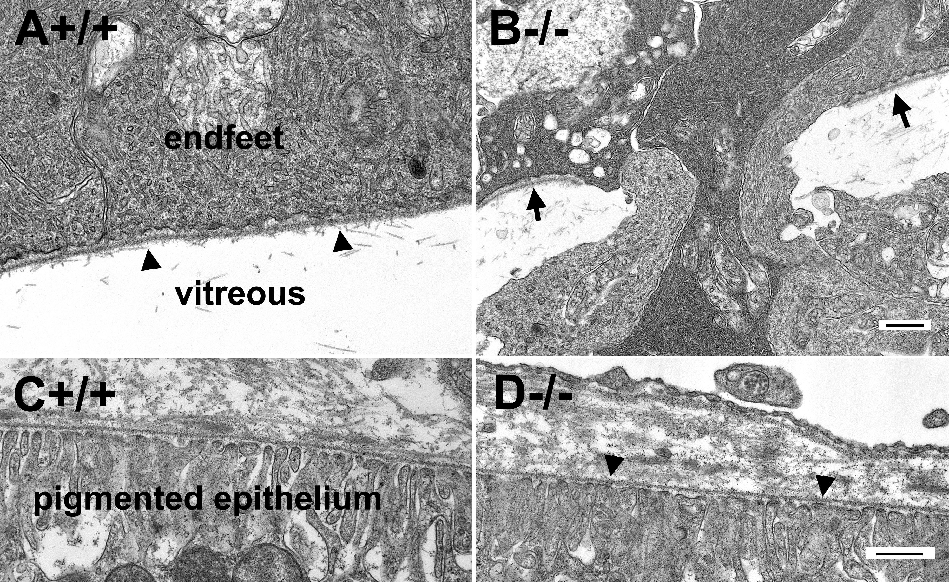
 Figure 2 of Hu, Mol Vis 2010; 16:1415-1428.