Figure 1. Dystroglycan distribution at the
Müller glia endfeet and inner limiting membrane are disrupted in
protein O-mannose N-acetylglucosaminyltransferase 1 (POMGnT1) knockout
retina. Adult retinal sections were double stained with
anti-β-dystroglycan (green fluorescence) and anti-laminin (red
fluorescence). Wild type (A-C) and knockout (D-F)
retinas
were immunostained with anti β-dystroglycan (A, D)
and laminin (B, E). All sections were counterstained
with 4',6-diamidino-2-phenylindole to show nuclei. Merged image of A
and B is shown in C. Merged image of D and E
is shown in F. Note β-dystroglycan and laminin staining at the
inner limiting membrane had breaks (arrows in D and E),
indicating a lack of Müller glia endfeet and inner limiting membrane at
these locations. Abbreviations: DG represents dystroglycan; ILM
represents inner limiting membrane; INL represents inner nuclear layer;
IPL represents inner plexiform layer; ONL represents outer nuclear
layer; OPL represents outer plexiform layer, DAPI represents
4',6-diamidino-2-phenylindole; +/+ represents wild type; −/− represents
homozygous knockout. Scale bar in F is equal to 50 μm.
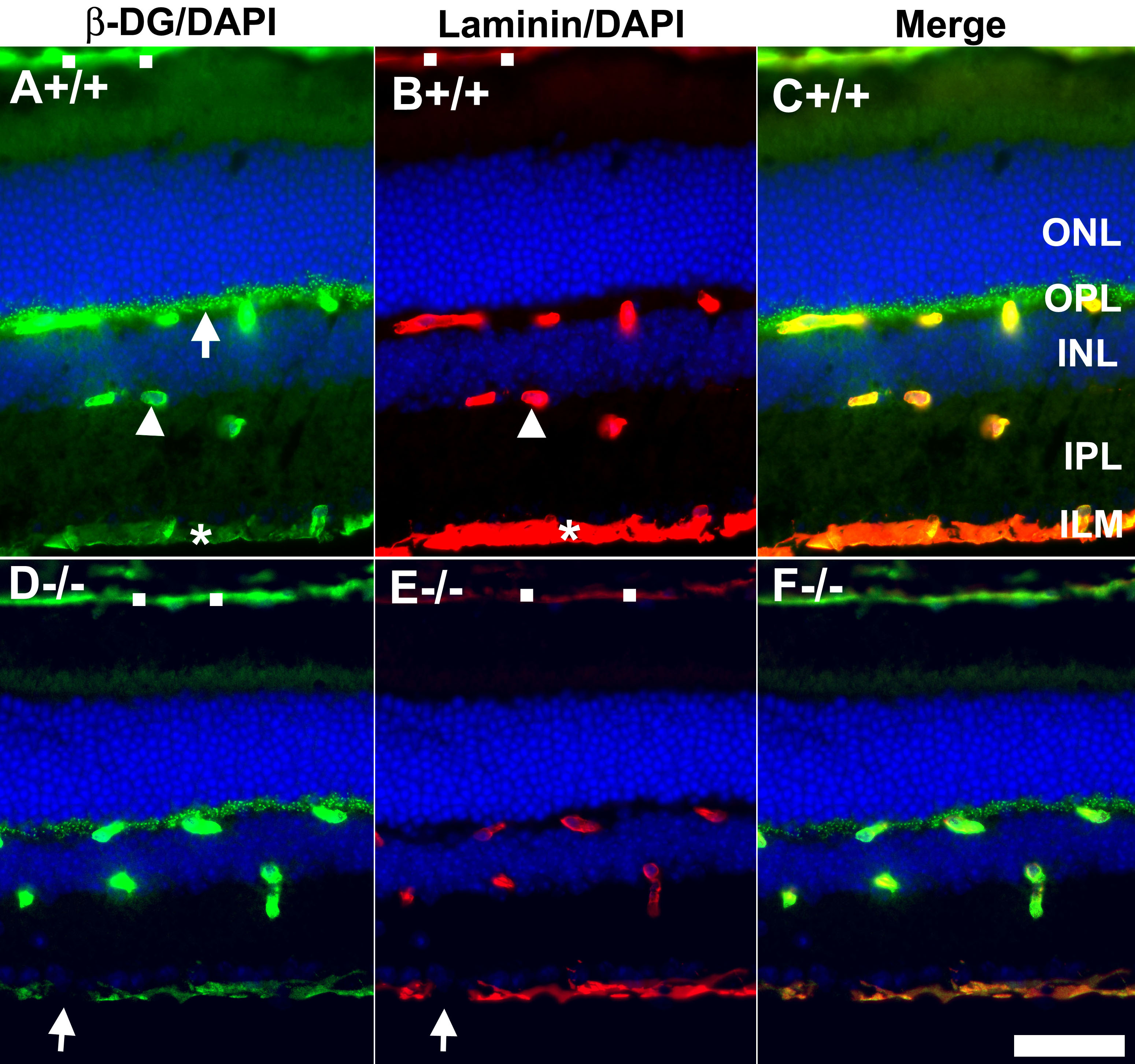
 Figure 1 of Hu, Mol Vis 2010; 16:1415-1428.