Figure 4. Immunofluorescence microscopy
analysis of p62 (red) and ubiquitin (green) in ARPE-19 cells. Cells
were exposed to 5 µM MG-132 with nonsilencing siRNA or with p62
siRNA (both 30 nM) for 24 h. Nuclei are stained with blue dye. Arrows
point to the colocalization of p62 and ubiquitin in perinuclear protein
aggregates.
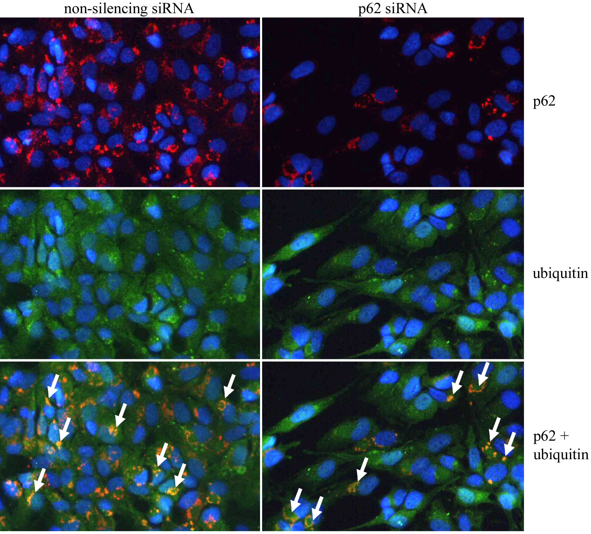
 Figure 4 of Viiri, Mol Vis 2010; 16:1399-1414.