Figure 4. Expression of vasohibin-1 protein within mouse cornea, as detected by western blot analysis. Mice were administered with subconjunctival
injection of Ad-Vasohibin-1 at 109 viral particles. Corneas were harvested at different time points, and protein samples were extracted. Ten corneal samples
were pooled together for each western blot analysis. A mouse antihuman vasohibin-1 monoclonal antibody was used for the western
blot. No vasohibin-1 western blot signal was detected before Ad-Vasohibin-1 was injected. A relatively weak signal was detected
on day 5 and was maintained at high levels until day 14 after injection.
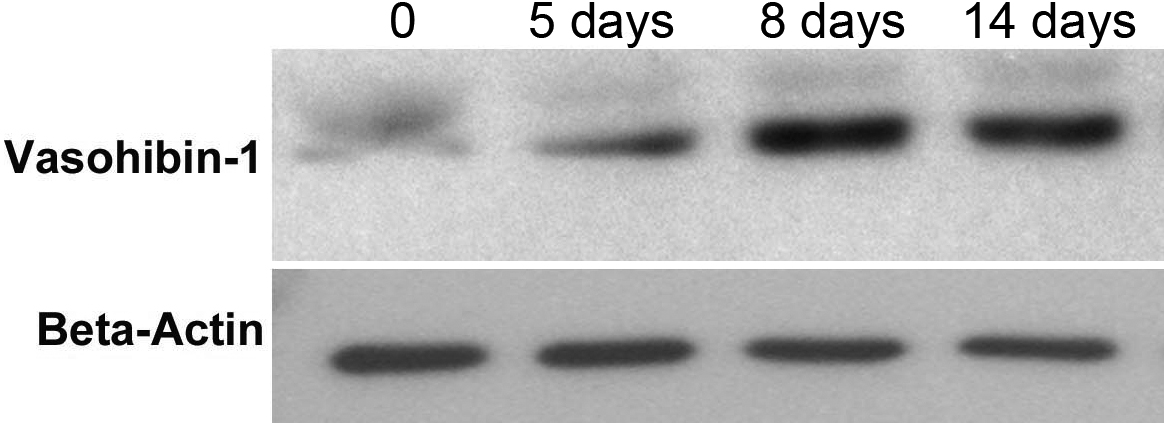
 Figure 4 of
Zhou, Mol Vis 2010; 16:1389-1398.
Figure 4 of
Zhou, Mol Vis 2010; 16:1389-1398.