Figure 3. The expression of exogenous
human vasohibin-1 protein in mouse corneas at different time points
after alkali-induced injury. Ad-Vasohibin-1 was injected
subconjunctivally 5 days before alkali-induced corneal injury at a
titer of 109 viral particles. A–C: On day 3
after alkali-induced injury (8 days after injection), vasohibin-1 was
expressed within the injection sites and some staining signals were
detected at the roots of the iris. D–F:On day 6 after
alkali treatment, vasohibin-1 was highly expressed in the subepithelial
stroma of the cornea, and some immunostaining was detected in the deep
stroma of the central cornea. Vasohibin-1 expression was highly
co-localized with corneal neovascularzation. Immunostaining was
diffusely distributed in the frontal stroma, which was not co-localized
with new blood vessels within the cornea. G–I: On day 9,
peripheral cornea showed a similar staining pattern as central cornea,
but more vasohibin-1 expression was detected in the deep corneal
stroma. J–L: There was no positive immunostaining for
vasohibin-1 and CD31 antigen in normal cornea without subconjunctival
injection.
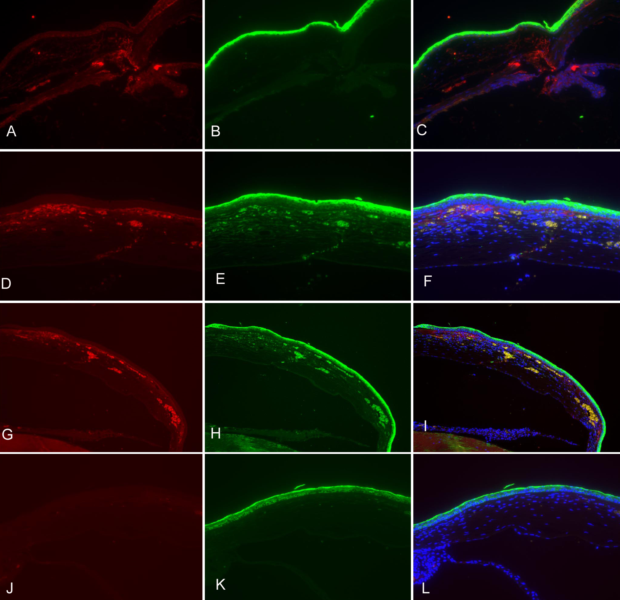
 Figure 3 of Zhou, Mol Vis 2010; 16:1389-1398.