Figure 1. The percentages of neovascularized cornea at each time point. The percentage of neovascularized cornea increased over time
in both groups but was significantly reduced in mice with subconjunctival injection of recombinant adenovirus encoding human
Vasohibin-1 gene (Ad-Vasohibin-1) compared with the blank adenoviral vector (AdNull) at all time points after alkali-induced
injury. On day 3, there was statistical difference in the percentage of CNV area between Ad-Vasohibin-1 (n=38) and Ad Null
(n=37) groups (F=50.26, t=–11.940, 95% CI of the difference: –9.78% to –6.80%). The same results were obtained on day 6 between Ad-Vasohibin-1 (n=25)
and Ad Null (n=24) groups (F=3.953, t=–7.868, 95% CI of the difference: –15.42% to –9.14%) and on day 9 between Ad-Vasohibin-1 (n=22) and Ad Null (n=21) groups
(F=2.318, t=–7.975, 95% CI of the difference: –26.60% to –15.85%). The asterisk indicates a p<0.01.
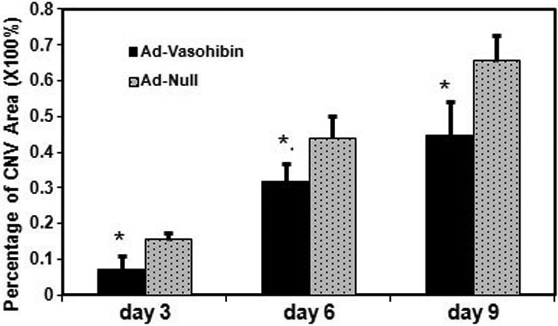
 Figure 1 of
Zhou, Mol Vis 2010; 16:1389-1398.
Figure 1 of
Zhou, Mol Vis 2010; 16:1389-1398.