Figure 1. Impact of EPA on firm leukocyte adhesion in the retinal vessels during EIU. A: Representative micrographs of flatmounted retinas from normal control mice (a,b), vehicle-treated EIU mice (c,d) and EPA-treated
(50 mg/kg BW) EIU mice (e,f) at 24 h after treatment and EIU-induction. Firmly adhering leukocytes in the retinal vasculature
were visualized by perfusion with ConA. Arrows indicate firmly adhering leukocytes in the inflamed retinal vasculature (c-e).
Scale bars=100 µm. B: Quantification of firm adhering leukocytes in the retinal vessels. Values are means±SEM (n=9 to 16). †p<0.01.
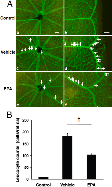
 Figure 1 of
Suzuki, Mol Vis 2010; 16:1382-1388.
Figure 1 of
Suzuki, Mol Vis 2010; 16:1382-1388.