Figure 3. Immunodepletion of NPHP4 in bovine retina. A: Schematic representation of the procedure used for ID/IP experiments. B: About 500 mg of bovine retinal lysate was subjected to IP using anti-NPHP4 antibody. Precipitated (pellet) as well as supernatant
samples were analyzed by immunoblotting using NPHP4 antibody. C: NPHP4-immunodepleted supernatant (NPHP4-ID sup) was subjected to IP with anti-RPGR antibody followed by immunoblot analysis
using the indicated antibodies. Molecular weight markers in kDa are shown on the left.
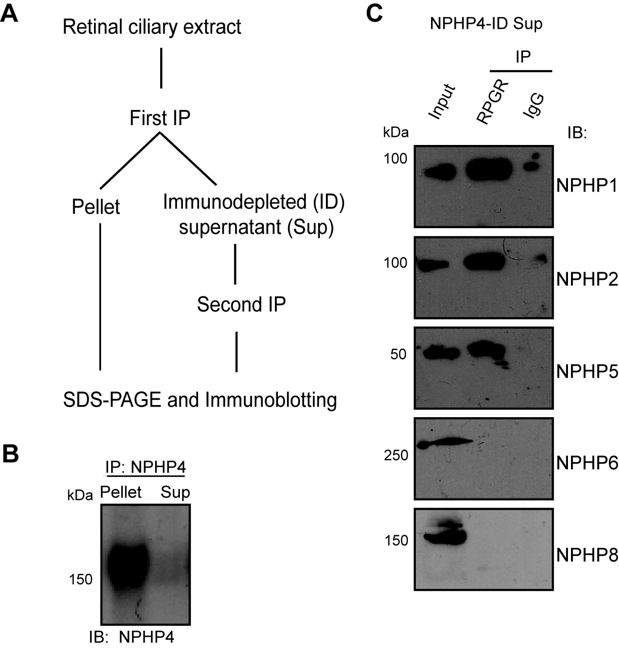
 Figure 3 of
Murga-Zamalloa, Mol Vis 2010; 16:1373-1381.
Figure 3 of
Murga-Zamalloa, Mol Vis 2010; 16:1373-1381.