Figure 2. RPGR interacts with NPHP1. A,
B: The association of RPGR with NPHP1 in bovine retina was
analyzed by co-IP using appropriate antibodies. Precipitated proteins
were analyzed by SDS–PAGE followed by immunoblotting with the indicated
antibodies. C: Schematic representation of the primary
structure of NPHP1. Abbreviations: NHD represents the nephrocystin
homology domain; BD represents the binding domain; SH3 represents the
Src homology domain. D: Interaction of GST-RPGR with 35S
NPHP1
was analyzed by GST pull-down assay. Amount of GST bound proteins
was evaluated by Coomassie blue staining (asterisk; lower panel). E:
This
panel denotes expression of different domains of 35S-NPHP1
(asterisks) used in the GST pull-down assay. Gels were analyzed by
autoradiography. F: The interaction of different domains of
NPHP1 with RPGR was analyzed by GST pull-down assay. Coomassie blue
staining of the gels loaded with GST-RPGR protein was performed to
evaluate the amount of protein in each experiment (lower panel).
Molecular weight markers in kDa are shown on the left. Arrows indicate
the specific protein bands representing the 35S-NPHP1
deleted domains.
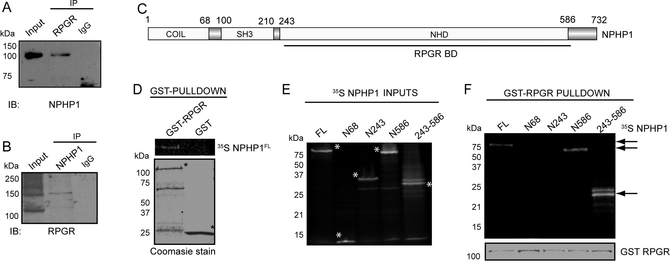
 Figure 2 of Murga-Zamalloa, Mol Vis 2010; 16:1373-1381.